
EFhEF-hand, calcium binding motif |
|---|
| SMART accession number: | SM00054 |
|---|---|
| Description: | EF-hands are calcium-binding motifs that occur at least in pairs. Links between disease states and genes encoding EF-hands, particularly the S100 subclass, are emerging. Each motif consists of a 12 residue loop flanked on either side by a 12 residue alpha-helix. EF-hands undergo a conformational change unpon binding calcium ions. |
| Interpro abstract (IPR002048): | Many calcium-binding proteins belong to the same evolutionary family and share a type of calcium-binding domain known as the EF-hand. This type of domain consists of a twelve residue loop flanked on both sides by a twelve residue alpha-helical domain. In an EF-hand loop the calcium ion is coordinated in a pentagonal bipyramidal configuration. The six residues involved in the binding are in positions 1, 3, 5, 7, 9 and 12; these residues are denoted by X, Y, Z, -Y, -X and -Z. The invariant Glu or Asp at position 12 provides two oxygens for liganding Ca (bidentate ligand). Ca2 binding induces a conformational change in the EF-hand motif, leading to the activation or inactivation of target proteins. EF-hands tend to occur in pairs or higher copy numbers [ (PUBMED:9228939) (PUBMED:8848832) (PUBMED:7553064) (PUBMED:7656053) (PUBMED:10591109) ]. |
| GO function: | calcium ion binding (GO:0005509) |
| Family alignment: |
There are 343947 EFh domains in 122495 proteins in SMART's nrdb database.
Click on the following links for more information.
- Evolution (species in which this domain is found)
-
Taxonomic distribution of proteins containing EFh domain.
This tree includes only several representative species. The complete taxonomic breakdown of all proteins with EFh domain is also avaliable.
Click on the protein counts, or double click on taxonomic names to display all proteins containing EFh domain in the selected taxonomic class.
- Cellular role (predicted cellular role)
-
Binding / catalysis: Ca2+-binding
- Literature (relevant references for this domain)
-
Primary literature is listed below; Automatically-derived, secondary literature is also avaliable.
- Kretsinger RH
- EF-hands embrace.
- Nat Struct Biol. 1997; 4: 514-6
- Ikura M
- Calcium binding and conformational response in EF-hand proteins.
- Trends Biochem Sci. 1996; 21: 14-7
- Display abstract
EF-hand proteins undergo conformational changes upon binding of Ca2+. This event is a crucial step in many Ca2+-dependent cellular processes. Recent advances in the three-dimensional structural analysis of various EF-hand proteins have led to new insights into the structure and functional relationship of this large family of Ca2+-binding proteins.
- Schafer BW, Heizmann CW
- The S100 family of EF-hand calcium-binding proteins: functions and pathology.
- Trends Biochem Sci. 1996; 21: 134-40
- Display abstract
Calcium lons as second messengers control many biological processes, at least in part, via interaction with a large number of Ca(2+)-binding proteins. One class of these proteins shares a common Ca(2+)-binding motif, the EF-hand, Here, we describe some functional aspects of EF-hand proteins, which have been found recently in different cellular compartments. Novel links between EF-hand proteins, particularly S100 proteins, and specific diseases are now emerging.
- Akke M, Forsen S, Chazin WJ
- Solution structure of (Cd2+)1-calbindin D9k reveals details of the stepwise structural changes along the Apo-->(Ca2+)II1-->(Ca2+)I,II2 binding pathway.
- J Mol Biol. 1995; 252: 102-21
- Display abstract
The three-dimensional solution structure of (Cd2+)1-calbindin D9k has been determined by distance geometry, restrained molecular dynamics and relaxation matrix calculations using experimental constraints obtained from two-dimensional 1H and 15N-1H NMR spectroscopy. The final input data consisted of 1055 NOE distance constraints and 71 dihedral angle constraints, corresponding to 15 constraints per residue on average. The resulting ensemble of 24 structures has no distance or dihedral angle constraints consistently violated by more than 0.07 A and 1.8 degrees, respectively. The structure is characteristic of an EF-hand protein, with two helix-loop-helix calcium binding motifs joined by a flexible linker, and a short anti-parallel beta-type interaction between the two ion-binding sites. The four helices are well defined with a root mean square deviation from the mean coordinates of 0.35 A for the backbone atoms. The structure of the half-saturated cadmium state was compared with the previously determined solution structures of the apo and fully calcium saturated calbindin D9k. The comparisons were aided by introducing the ensemble averaged distance difference matrix as a tool for analyzing differences between two ensembles of structures. Detailed analyses of differences between the three states in backbone and side-chain dihedral angles, hydrogen bonds, interatomic distances, and packing of the hydrophobic core reveal the reorganization of the protein that occurs upon ion binding. Overall, it was found that (Cd2+)1-calbindin D9k, representing the half-saturated calcium state with an ion in site II, is structurally more similar to the fully calcium-saturated state than the apo state. Thus, for the binding sequence apo-->(Ca2+)II1-->(Ca2+)I,II2, the structural changes occurring upon ion binding are most pronounced for the first binding step, an observation that bears significantly on the molecular basis for cooperative calcium binding in calbindin D9k.
- Kawasaki H, Kretsinger RH
- Calcium-binding proteins 1: EF-hands.
- Protein Profile. 1995; 2: 297-490
- Potts BC et al.
- The structure of calcyclin reveals a novel homodimeric fold for S100 Ca(2+)-binding proteins.
- Nat Struct Biol. 1995; 2: 790-6
- Display abstract
The S100 calcium-binding proteins are implicated as effectors in calcium-mediated signal transduction pathways. The three-dimensional structure of the S100 protein calcyclin has been determined in solution in the apo state by NMR spectroscopy and a computational strategy that incorporates a systematic docking protocol. This structure reveals a symmetric homodimeric fold that is unique among calcium-binding proteins. Dimerization is mediated by hydrophobic contacts from several highly conserved residues, which suggests that the dimer fold identified for calcyclin will serve as a structural paradigm for the S100 subfamily of calcium-binding proteins.
- Skelton NJ, Kordel J, Chazin WJ
- Determination of the solution structure of Apo calbindin D9k by NMR spectroscopy.
- J Mol Biol. 1995; 249: 441-62
- Display abstract
The three-dimensional structure of apo calbindin D9k has been determined using constraints generated from nuclear magnetic resonance spectroscopy. The family of solution structures was calculated using a combination of distance geometry, restrained molecular dynamics, and hybrid relaxation matrix analysis of the nuclear Overhauser effect (NOE) cross-peak intensities. Errors and inconsistencies in the input constraints were identified using complete relaxation matrix analyses based on the results of preliminary structure calculations. The final input data consisted of 994 NOE distance constraints and 122 dihedral constraints, aided by the stereospecific assignment of the resonances from 21 beta-methylene groups and seven isopropyl groups of leucine and valine residues. The resulting family of 33 structures contain no violation of the distance constraints greater than 0.17 A or of the dihedral angle constraints greater than 10 degrees. The structures consist of a well-defined, antiparallel four-helix bundle, with a short anti-parallel beta-interaction between the two unoccupied calcium-binding loops. The root-mean-square deviation from the mean structure of the backbone heavy-atoms for the well-defined helical residues is 0.55 A. The remainder of the ion-binding loops, the linker loop connecting the two sub-domains of the protein, and the N and C termini exhibit considerable disorder between different structures in the ensemble. A comparison with the structure of the (Ca2+)2 state indicates that the largest changes associated with ion-binding occur in the middle of helix IV and in the packing of helix III onto the remainder of the protein. The change in conformation of these helices is associated with a subtle reorganization of many residues in the hydrophobic core, including some side-chains that are up to 15 A from the ion-binding site.
- Slupsky CM, Kay CM, Reinach FC, Smillie LB, Sykes BD
- Calcium-induced dimerization of troponin C: mode of interaction and use of trifluoroethanol as a denaturant of quaternary structure.
- Biochemistry. 1995; 34: 7365-75
- Display abstract
Protein aggregation can be a problem, especially as a large number of proteins become available for structural studies at fairly high concentrations using solution techniques such as NMR spectroscopy. The muscle regulatory protein troponin C (TnC) undergoes a calcium-induced dimerization at neutral pH with a dissociation constant for the dimerization of 0.4 mM at 20 degrees C. The present study indicates that the mode of dimerization involves the N-domain of one monomer interacting with the N-domain of another monomer. Addition of the solvent trifluoroethanol (TFE) to a concentration of 15%, v/v, results in a 10-fold increase in the dimer dissociation constant of calcium-saturated TnC (4 mM at 20 degrees C), making TnC predominantly a monomer for spectroscopic studies. Further, TFE, at the concentrations used herein, acts to perturb the quaternary structure of TnC without adversely affecting the secondary or tertiary structure as evidenced by minimal changes to its CD spectra and 1H, 13C, and 15N NMR chemical shifts.
- Slupsky CM, Reinach FC, Smillie LB, Sykes BD
- Solution secondary structure of calcium-saturated troponin C monomer determined by multidimensional heteronuclear NMR spectroscopy.
- Protein Sci. 1995; 4: 1279-90
- Display abstract
The solution secondary structure of calcium-saturated skeletal troponin C (TnC) in the presence of 15% (v/v) trifluoroethanol (TFE), which has been shown to exist predominantly as a monomer (Slupsky CM, Kay CM, Reinach FC, Smillie LB, Sykes BD, 1995, Biochemistry 34, forthcoming), has been investigated using multidimensional heteronuclear nuclear magnetic resonance spectroscopy. The 1H, 15N, and 13C NMR chemical shift values for TnC in the presence of TFE are very similar to values obtained for calcium-saturated NTnC (residues 1-90 of skeletal TnC), calmodulin, and synthetic peptide homodimers. Moreover, the secondary structure elements of TnC are virtually identical to those obtained for calcium-saturated NTnC, calmodulin, and the synthetic peptide homodimers, suggesting that 15% (v/v) TFE minimally perturbs the secondary and tertiary structure of this stably folded protein. Comparison of the solution structure of calcium-saturated TnC with the X-ray crystal structure of half-saturated TnC reveals differences in the phi/psi angles of residue Glu 41 and in the linker between the two domains. Glu 41 has irregular phi/psi angles in the crystal structure, producing a kink in the B helix, whereas in calcium-saturated TnC, Glu 41 has helical phi/psi angles, resulting in a straight B helix. The linker between the N and C domains of calcium-saturated TnC is flexible in the solution structure.
- Gagne SM, Tsuda S, Li MX, Chandra M, Smillie LB, Sykes BD
- Quantification of the calcium-induced secondary structural changes in the regulatory domain of troponin-C.
- Protein Sci. 1994; 3: 1961-74
- Display abstract
The backbone resonance assignments have been completed for the apo (1H and 15N) and calcium-loaded (1H, 15N, and 13C) regulatory N-domain of chicken skeletal troponin-C (1-90), using multidimensional homonuclear and heteronuclear NMR spectroscopy. The chemical-shift information, along with detailed NOE analysis and 3JHNH alpha coupling constants, permitted the determination and quantification of the Ca(2+)-induced secondary structural change in the N-domain of TnC. For both structures, 5 helices and 2 short beta-strands were found, as was observed in the apo N-domain of the crystal structure of whole TnC (Herzberg O, James MNG, 1988, J Mol Biol 203:761-779). The NMR solution structure of the apo form is indistinguishable from the crystal structure, whereas some structural differences are evident when comparing the 2Ca2+ state solution structure with the apo one. The major conformational change observed is the straightening of helix-B upon Ca2+ binding. The possible importance and role of this conformational change is explored. Previous CD studies on the regulatory domain of TnC showed a significant Ca(2+)-induced increase in negative ellipticity, suggesting a significant increase in helical content upon Ca2+ binding. The present study shows that there is virtually no change in alpha-helical content associated with the transition from apo to the 2Ca2+ state of the N-domain of TnC. Therefore, the Ca(2+)-induced increase in ellipticity observed by CD does not relate to a change in helical content, but more likely to changes in spatial orientation of helices.
- Skelton NJ, Kordel J, Akke M, Forsen S, Chazin WJ
- Signal transduction versus buffering activity in Ca(2+)-binding proteins.
- Nat Struct Biol. 1994; 1: 239-45
- Display abstract
The three-dimensional structure of calbindin D9k in the absence of Ca2+ has been determined using NMR spectroscopy in solution, allowing the first direct analysis of the consequences of Ca2+ binding for a member of the calmodulin superfamily of proteins. The overall response in calbindin D9k is much attenuated relative to the current model for calmodulin and troponin C. These results demonstrate a novel mechanism for modulating the conformational response to Ca(2+)-binding in calmodulin superfamily proteins and provide insights into how their Ca(2+)-binding domains can be fine-tuned to remain essentially intact or respond strongly to ion binding, in relation to their functional requirements.
- Kordel J, Skelton NJ, Akke M, Chazin WJ
- High-resolution structure of calcium-loaded calbindin D9k.
- J Mol Biol. 1993; 231: 711-34
- Display abstract
The three-dimensional solution structure of calcium-loaded calbindin D9k has been determined using experimental constraints obtained from nuclear magnetic resonance spectroscopy. A total of 1176 constraints (16 per residue overall, 32 per residue for the core residues) was used for the final refinement, including 1002 distance and 174 dihedral angle constraints. In addition, 23 hydrogen bond constraints were used for the generation of initial structures. Stereospecific assignments were made for 37 of 61 (61%) prochiral methylene protons and the methyl groups of all three valine residues and five out of 12 leucine residues. These constraints were used as input for a series of calculations of three-dimensional structures using a combination of distance geometry and restrained molecular dynamics. The 33 best structures selected for further analysis have no distance constraint violations greater than 0.3 A and good local geometries as reflected by low total energies (< or = -1014 kcal/mol in the AMBER 4.0 force field). The core of the protein consists of four well-defined helices with root-mean-square deviations from the average of 0.45 A for the N, C alpha and C' backbone atoms. These helices are packed in an antiparallel fashion to form two helix-loop-helix calcium-binding motifs, termed EF-hands. The two EF-hands are joined at one end by a ten-residue linker segment, and at the other by a short beta-type interaction between the two calcium-binding loops. Overall, the average solution structure of calbindin D9k is very similar to the crystal structure, with a pairwise root-mean-square deviation of 0.85 A for the N, C alpha and C' backbone atoms of the four helices. The differences that are observed between the solution and the crystal structures are attributed to specific crystal contacts, increased side-chain flexibility in solution, or artifacts arising from molecular dynamics refinement of the solution structures in vacuo.
- Watterson DM, HarrelsonWGJ r, Keller PM, Sharief F, Vanaman TC
- Structural similarities between the Ca2+-dependent regulatory proteins of 3':5'-cyclic nucleotide phosphodiesterase and actomyosin ATPase.
- J Biol Chem. 1976; 251: 4501-13
- Display abstract
Results of studies of the Ca2+-dependent protein modulator of 3':5'-cyclic nucleotide phosphodiesterase isolated from bovine brain are presented which show its structural similarity to the Ca2+-binding subunit of muscle troponin. Both proteins have blocked NH2 termini, similar and characteristic ultraviolet absorption spectra, similar Ca2+-binding properties, very similar amino acid compositions, and co-migrate on sodium dodecyl sulfate-polyacrylamide gels. The primary structures of selected tryptic peptides isolated from bovine brain modulator protein are similar or identical with regions of the primary sequences of rabbit skeletal muscle and bovine cardiac muscle troponin C. Bovine brain modulator protein contains and unidentified ninhydrin-positive basic compound not found in muscle troponin C. An improved procedure is presented which yields 40 to 70 mg of modulator protein per kg of bovine brain.
- Kretsinger RH
- Gene triplication deduced from the tertiary structure of a muscle calcium binding protein.
- Nat New Biol. 1972; 240: 85-8
- McLachlan AD
- Gene duplication in carp muscle calcium binding protein.
- Nat New Biol. 1972; 240: 83-5
- Disease (disease genes where sequence variants are found in this domain)
-
SwissProt sequences and OMIM curated human diseases associated with missense mutations within the EFh domain.
Protein Disease Calpain-3 (P20807) (SMART) OMIM:114240: Muscular dystrophy, limb-girdle, type 2A
OMIM:253600: - Metabolism (metabolic pathways involving proteins which contain this domain)
-
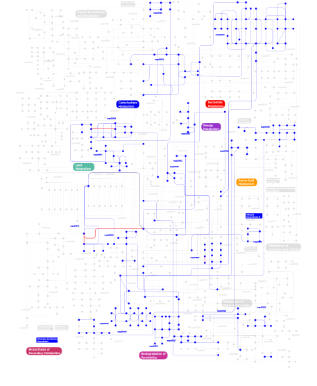
Click the image to view the interactive version of the map in iPath% proteins involved KEGG pathway ID Description 8.58 map04020 Calcium signaling pathway 7.04 map04510 Focal adhesion 6.27 map04810 Regulation of actin cytoskeleton 6.27 map04530 Tight junction 6.01 map04070 Phosphatidylinositol signaling system 5.41 map04720 Long-term potentiation 4.72 map04740 Olfactory transduction 4.12 map04210 Apoptosis 3.52 map04010 MAPK signaling pathway 3.26 map04670 Leukocyte transendothelial migration 3.00 map04520 Adherens junction 2.92 map05214 Glioma 2.92 map04916 Melanogenesis 2.92 map04912 GnRH signaling pathway 2.92 map04910 Insulin signaling pathway 2.92 map05040 Huntington's disease 2.83  map00562
Inositol phosphate metabolism 2.75 map00564Glycerophospholipid metabolism 2.49 map04370 VEGF signaling pathway 2.49 map04662 B cell receptor signaling pathway 2.49 map04310 Wnt signaling pathway 2.49 map04360 Axon guidance 2.49 map04660 T cell receptor signaling pathway 2.49 map04650 Natural killer cell mediated cytotoxicity 1.72 map00561Glycerolipid metabolism 1.63 map00632Benzoate degradation via CoA ligation 0.77 map00030Pentose phosphate pathway 0.26 map00190Oxidative phosphorylation 0.26 map00360Phenylalanine metabolism 0.17 map00350Tyrosine metabolism 0.17 map00340Histidine metabolism 0.17 map00642Ethylbenzene degradation 0.17 map00903 Limonene and pinene degradation 0.17 map00960 Alkaloid biosynthesis II 0.17 map00310Lysine degradation 0.17 map006241- and 2-Methylnaphthalene degradation 0.17 map00230Purine metabolism 0.09 map00940Phenylpropanoid biosynthesis 0.09 map00650Butanoate metabolism 0.09 map00680Methane metabolism 0.09 map00280Valine, leucine and isoleucine degradation 0.09 map00072Synthesis and degradation of ketone bodies 0.09 map00361gamma-Hexachlorocyclohexane degradation 0.09 map006271,4-Dichlorobenzene degradation 0.09 map00364 Fluorobenzoate degradation This information is based on mapping of SMART genomic protein database to KEGG orthologous groups. Percentage points are related to the number of proteins with EFh domain which could be assigned to a KEGG orthologous group, and not all proteins containing EFh domain. Please note that proteins can be included in multiple pathways, ie. the numbers above will not always add up to 100%.
- Structure (3D structures containing this domain)
3D Structures of EFh domains in PDB
PDB code Main view Title 1a29 
CALMODULIN COMPLEXED WITH TRIFLUOPERAZINE (1:2 COMPLEX) 1a2x 
COMPLEX OF TROPONIN C WITH A 47 RESIDUE (1-47) FRAGMENT OF TROPONIN I 1a75 
WHITING PARVALBUMIN 1ahr 
CALMODULIN MUTANT WITH A TWO RESIDUE DELETION IN THE CENTRAL HELIX 1aj4 
STRUCTURE OF CALCIUM-SATURATED CARDIAC TROPONIN C, NMR, 1 STRUCTURE 1aj5 
CALPAIN DOMAIN VI APO 1ak8 
NMR SOLUTION STRUCTURE OF CERIUM-LOADED CALMODULIN AMINO-TERMINAL DOMAIN (CE2-TR1C), 23 STRUCTURES 1alv 
CALCIUM BOUND DOMAIN VI OF PORCINE CALPAIN 1alw 
INHIBITOR AND CALCIUM BOUND DOMAIN VI OF PORCINE CALPAIN 1ap4 
REGULATORY DOMAIN OF HUMAN CARDIAC TROPONIN C IN THE CALCIUM-SATURATED STATE, NMR, 40 STRUCTURES 1aui 
HUMAN CALCINEURIN HETERODIMER 1avs 
X-RAY CRYSTALLOGRAPHIC STUDY OF CALCIUM-SATURATED N-TERMINAL DOMAIN OF TROPONIN C 1b7t 
MYOSIN DIGESTED BY PAPAIN 1b8r 
PARVALBUMIN 1b9a 
PARVALBUMIN (MUTATION;D51A, F102W) 1bjf 
CRYSTAL STRUCTURE OF RECOMBINANT BOVINE NEUROCALCIN DELTA AT 2.4 ANGSTROMS 1blq 
STRUCTURE AND INTERACTION SITE OF THE REGULATORY DOMAIN OF TROPONIN-C WHEN COMPLEXED WITH THE 96-148 REGION OF TROPONIN-I, NMR, 29 STRUCTURES 1boc 
THE SOLUTION STRUCTURES OF MUTANT CALBINDIN D9K'S, AS DETERMINED BY NMR, SHOW THAT THE CALCIUM BINDING SITE CAN ADOPT DIFFERENT FOLDS 1bod 
THE SOLUTION STRUCTURES OF MUTANT CALBINDIN D9K'S, AS DETERMINED BY NMR, SHOW THAT THE CALCIUM BINDING SITE CAN ADOPT DIFFERENT FOLDS 1br1 
SMOOTH MUSCLE MYOSIN MOTOR DOMAIN-ESSENTIAL LIGHT CHAIN COMPLEX WITH MGADP.ALF4 BOUND AT THE ACTIVE SITE 1br4 
SMOOTH MUSCLE MYOSIN MOTOR DOMAIN-ESSENTIAL LIGHT CHAIN COMPLEX WITH MGADP.BEF3 BOUND AT THE ACTIVE SITE 1bu3 
REFINED CRYSTAL STRUCTURE OF CALCIUM-BOUND SILVER HAKE (PI 4.2) PARVALBUMIN AT 1.65 A. 1c7v 
NMR SOLUTION STRUCTURE OF THE CALCIUM-BOUND C-TERMINAL DOMAIN (W81-S161) OF CALCIUM VECTOR PROTEIN FROM AMPHIOXUS 1c7w 
NMR SOLUTION STRUCTURE OF THE CALCIUM-BOUND C-TERMINAL DOMAIN (W81-S161) OF CALCIUM VECTOR PROTEIN FROM AMPHIOXUS 1cdl 
TARGET ENZYME RECOGNITION BY CALMODULIN: 2.4 ANGSTROMS STRUCTURE OF A CALMODULIN-PEPTIDE COMPLEX 1cdm 
MODULATION OF CALMODULIN PLASTICITY IN MOLECULAR RECOGNITION ON THE BASIS OF X-RAY STRUCTURES 1cdp 
RESTRAINED LEAST SQUARES REFINEMENT OF NATIVE (CALCIUM) AND CADMIUM-SUBSTITUTED CARP PARVALBUMIN USING X-RAY CRYSTALLOGRAPHIC DATA AT 1.6-ANGSTROMS RESOLUTION 1cfc 
CALCIUM-FREE CALMODULIN 1cfd 
CALCIUM-FREE CALMODULIN 1cff 
NMR SOLUTION STRUCTURE OF A COMPLEX OF CALMODULIN WITH A BINDING PEPTIDE OF THE CA2+-PUMP 1ckk 
CALMODULIN/RAT CA2+/CALMODULIN DEPENDENT PROTEIN KINASE FRAGMENT 1cll 
CALMODULIN STRUCTURE REFINED AT 1.7 ANGSTROMS RESOLUTION 1clm 
STRUCTURE OF PARAMECIUM TETRAURELIA CALMODULIN AT 1.8 ANGSTROMS RESOLUTION 1cm1 
MOTIONS OF CALMODULIN-SINGLE-CONFORMER REFINEMENT 1cm4 
MOTIONS OF CALMODULIN-FOUR-CONFORMER REFINEMENT 1cmf 
NMR SOLUTION STRUCTURE OF APO CALMODULIN CARBOXY-TERMINAL DOMAIN 1cmg 
NMR SOLUTION STRUCTURE OF CALCIUM-LOADED CALMODULIN CARBOXY-TERMINAL DOMAIN 1ctr 
DRUG BINDING BY CALMODULIN: CRYSTAL STRUCTURE OF A CALMODULIN-TRIFLUOPERAZINE COMPLEX 1deg 
THE LINKER OF DES-GLU84 CALMODULIN IS BENT AS SEEN IN THE CRYSTAL STRUCTURE 1df0 
Crystal structure of M-Calpain 1dfk 
NUCLEOTIDE-FREE SCALLOP MYOSIN S1-NEAR RIGOR STATE 1dfl 
SCALLOP MYOSIN S1 COMPLEXED WITH MGADP:VANADATE-TRANSITION STATE 1dgu 
HOMOLOGY-BASED MODEL OF CALCIUM-SATURATED CIB (CALCIUM-AND INTEGRIN-BINDING PROTEIN) 1dgv 
HOMOLOGY-BASED MODEL OF APO CIB (CALCIUM-AND INTEGRIN-BINDING PROTEIN) 1djg 
PHOSPHOINOSITIDE-SPECIFIC PHOSPHOLIPASE C-DELTA1 FROM RAT COMPLEXED WITH LANTHANUM 1djh 
PHOSPHOINOSITIDE-SPECIFIC PHOSPHOLIPASE C-DELTA1 FROM RAT COMPLEXED WITH BARIUM 1dji 
PHOSPHOINOSITIDE-SPECIFIC PHOSPHOLIPASE C-DELTA1 FROM RAT COMPLEXED WITH CALCIUM 1djw 
PHOSPHOINOSITIDE-SPECIFIC PHOSPHOLIPASE C-DELTA1 FROM RAT COMPLEXED WITH INOSITOL-2-METHYLENE-1,2-CYCLIC-MONOPHOSPHONATE 1djx 
PHOSPHOINOSITIDE-SPECIFIC PHOSPHOLIPASE C-DELTA1 FROM RAT COMPLEXED WITH INOSITOL-1,4,5-TRISPHOSPHATE 1djy 
PHOSPHOINOSITIDE-SPECIFIC PHOSPHOLIPASE C-DELTA1 FROM RAT COMPLEXED WITH INOSITOL-2,4,5-TRISPHOSPHATE 1djz 
PHOSPHOINOSITIDE-SPECIFIC PHOSPHOLIPASE C-DELTA1 FROM RAT COMPLEXED WITH INOSITOL-4,5-BISPHOSPHATE 1dmo 
CALMODULIN, NMR, 30 STRUCTURES 1dtl 
CRYSTAL STRUCTURE OF CALCIUM-SATURATED (3CA2+) CARDIAC TROPONIN C COMPLEXED WITH THE CALCIUM SENSITIZER BEPRIDIL AT 2.15 A RESOLUTION 1dvi 
CALPAIN DOMAIN VI WITH CALCIUM BOUND 1ej3 
CRYSTAL STRUCTURE OF AEQUORIN 1el4 
STRUCTURE OF THE CALCIUM-REGULATED PHOTOPROTEIN OBELIN DETERMINED BY SULFUR SAS 1exr 
THE 1.0 ANGSTROM CRYSTAL STRUCTURE OF CA+2 BOUND CALMODULIN 1f4o 
CRYSTAL STRUCTURE OF GRANCALCIN WITH BOUND CALCIUM 1f4q 
CRYSTAL STRUCTURE OF APO GRANCALCIN 1f54 
SOLUTION STRUCTURE OF THE APO N-TERMINAL DOMAIN OF YEAST CALMODULIN 1f55 
SOLUTION STRUCTURE OF THE CALCIUM BOUND N-TERMINAL DOMAIN OF YEAST CALMODULIN 1f70 
REFINED SOLUTION STRUCTURE OF CALMODULIN N-TERMINAL DOMAIN 1f71 
REFINED SOLUTION STRUCTURE OF CALMODULIN C-TERMINAL DOMAIN 1fi5 
NMR STRUCTURE OF THE C TERMINAL DOMAIN OF CARDIAC TROPONIN C BOUND TO THE N TERMINAL DOMAIN OF CARDIAC TROPONIN I. 1fpw 
STRUCTURE OF YEAST FREQUENIN 1fw4 
CRYSTAL STRUCTURE OF E. COLI FRAGMENT TR2C FROM CALMODULIN TO 1.7 A RESOLUTION 1g33 
CRYSTAL STRUCTURE OF RAT PARVALBUMIN WITHOUT THE N-TERMINAL DOMAIN 1g4y 
1.60 A CRYSTAL STRUCTURE OF THE GATING DOMAIN FROM SMALL CONDUCTANCE POTASSIUM CHANNEL COMPLEXED WITH CALCIUM-CALMODULIN 1g8i 
CRYSTAL STRUCTURE OF HUMAN FREQUENIN (NEURONAL CALCIUM SENSOR 1) 1ggw 
CDC4P FROM SCHIZOSACCHAROMYCES POMBE 1ggz 
CRYSTAL STRUCTURE OF THE CALMODULIN-LIKE PROTEIN (HCLP) FROM HUMAN EPITHELIAL CELLS 1gjy 
The X-ray structure of the Sorcin Calcium Binding Domain (SCBD) provides insight into the phosphorylation and calcium dependent processess 1h4b 
SOLUTION STRUCTURE OF THE BIRCH POLLEN ALLERGEN BET V 4 1hqv 
STRUCTURE OF APOPTOSIS-LINKED PROTEIN ALG-2 1i84 
CRYO-EM STRUCTURE OF THE HEAVY MEROMYOSIN SUBFRAGMENT OF CHICKEN GIZZARD SMOOTH MUSCLE MYOSIN WITH REGULATORY LIGHT CHAIN IN THE DEPHOSPHORYLATED STATE. ONLY C ALPHAS PROVIDED FOR REGULATORY LIGHT CHAIN. ONLY BACKBONE ATOMS PROVIDED FOR S2 FRAGMENT. 1ih0 
Structure of the C-domain of Human Cardiac Troponin C in Complex with Ca2+ Sensitizer EMD 57033 1ij5 
METAL-FREE STRUCTURE OF MULTIDOMAIN EF-HAND PROTEIN, CBP40, FROM TRUE SLIME MOLD 1ij6 
CA2+-BOUND STRUCTURE OF MULTIDOMAIN EF-HAND PROTEIN, CBP40, FROM TRUE SLIME MOLD 1iku 
MYRISTOYLATED RECOVERIN IN THE CALCIUM-FREE STATE, NMR, 22 STRUCTURES 1iq5 
Calmodulin/nematode CA2+/Calmodulin dependent kinase kinase fragment 1iwq 
Crystal Structure of MARCKS calmodulin binding domain peptide complexed with Ca2+/Calmodulin 1j1d 
Crystal structure of the 46kDa domain of human cardiac troponin in the Ca2+ saturated form 1j1e 
Crystal structure of the 52kDa domain of human cardiac troponin in the Ca2+ saturated form 1j7o 
Solution structure of Calcium-calmodulin N-terminal domain 1j7p 
Solution structure of Calcium calmodulin C-terminal domain 1jba 
UNMYRISTOYLATED GCAP-2 WITH THREE CALCIUM IONS BOUND 1jc2 
COMPLEX OF THE C-DOMAIN OF TROPONIN C WITH RESIDUES 1-40 OF TROPONIN I 1jf0 
The Crystal Structure of Obelin from Obelia geniculata at 1.82 A Resolution 1jf2 
Crystal Structure of W92F obelin mutant from Obelia longissima at 1.72 Angstrom resolution 1jfj 
NMR SOLUTION STRUCTURE OF AN EF-HAND CALCIUM BINDING PROTEIN FROM ENTAMOEBA HISTOLYTICA 1jfk 
MINIMUM ENERGY REPRESENTATIVE STRUCTURE OF A CALCIUM BOUND EF-HAND PROTEIN FROM ENTAMOEBA HISTOLYTICA 1jsa 
MYRISTOYLATED RECOVERIN WITH TWO CALCIUMS BOUND, NMR, 24 STRUCTURES 1juo 
Crystal Structure of Calcium-free Human Sorcin: A Member of the Penta-EF-Hand Protein Family 1k90 
Crystal structure of the adenylyl cyclase domain of anthrax edema factor (EF) in complex with calmodulin and 3' deoxy-ATP 1k93 
Crystal structure of the adenylyl cyclase domain of anthrax edema factor (EF) in complex with calmodulin 1k94 
Crystal structure of des(1-52)grancalcin with bound calcium 1k95 
Crystal structure of des(1-52)grancalcin with bound calcium 1k9u 
Crystal Structure of the Calcium-Binding Pollen Allergen Phl p 7 (Polcalcin) at 1.75 Angstroem 1kfu 
Crystal Structure of Human m-Calpain Form II 1kfx 
Crystal Structure of Human m-Calpain Form I 1kk7 
SCALLOP MYOSIN IN THE NEAR RIGOR CONFORMATION 1kk8 
SCALLOP MYOSIN (S1-ADP-BeFx) IN THE ACTIN-DETACHED CONFORMATION 1kqm 
SCALLOP MYOSIN S1-AMPPNP IN THE ACTIN-DETACHED CONFORMATION 1kwo 
SCALLOP MYOSIN S1-ATPgammaS-p-PDM IN THE ACTIN-DETACHED CONFORMATION 1l2o 
SCALLOP MYOSIN S1-ADP-p-PDM IN THE ACTIN-DETACHED CONFORMATION 1l7z 
Crystal structure of Ca2+/Calmodulin complexed with myristoylated CAP-23/NAP-22 peptide 1la0 
Solution Structure of Calcium Saturated Cardiac Troponin C in the Troponin C-Troponin I Complex 1la3 
Solution structure of recoverin mutant, E85Q 1lin 
CALMODULIN COMPLEXED WITH TRIFLUOPERAZINE (1:4 COMPLEX) 1lkj 
NMR Structure of Apo Calmodulin from Yeast Saccharomyces cerevisiae 1lvc 
Crystal structure of the adenylyl cyclase domain of anthrax edema factor (EF) in complex with calmodulin and 2' deoxy, 3' anthraniloyl ATP 1lxf 
Structure of the Regulatory N-domain of Human Cardiac Troponin C in Complex with Human Cardiac Troponin-I(147-163) and Bepridil 1m39 
Solution structure of the C-terminal fragment (F86-I165) of the human centrin 2 in calcium saturated form 1m45 
CRYSTAL STRUCTURE OF MLC1P BOUND TO IQ2 OF MYO2P, A CLASS V MYOSIN 1m46 
CRYSTAL STRUCTURE OF MLC1P BOUND TO IQ4 OF MYO2P, A CLASS V MYOSIN 1m63 
Crystal structure of calcineurin-cyclophilin-cyclosporin shows common but distinct recognition of immunophilin-drug complexes 1m8q 
Molecular Models of Averaged Rigor Crossbridges from Tomograms of Insect Flight Muscle 1mf8 
Crystal Structure of human calcineurin complexed with cyclosporin A and human cyclophilin 1mux 
SOLUTION NMR STRUCTURE OF CALMODULIN/W-7 COMPLEX: THE BASIS OF DIVERSITY IN MOLECULAR RECOGNITION, 30 STRUCTURES 1mvw 
MOLECULAR MODELS OF AVERAGED RIGOR CROSSBRIDGES FROM TOMOGRAMS OF INSECT FLIGHT MUSCLE 1mxe 
Structure of the Complex of Calmodulin with the Target Sequence of CaMKI 1mxl 
STRUCTURE OF CARDIAC TROPONIN C-TROPONIN I COMPLEX 1n0y 
Crystal Structure of Pb-bound Calmodulin 1n2d 
Ternary complex of MLC1P bound to IQ2 and IQ3 of Myo2p, a class V myosin 1ncx 
TROPONIN C 1ncy 
TROPONIN-C, COMPLEX WITH MANGANESE 1ncz 
TROPONIN C 1niw 
Crystal structure of endothelial nitric oxide synthase peptide bound to calmodulin 1np8 
18-k C-terminally trunucated small subunit of calpain 1npq 
structure of a rhodamine-labeled N-domain Troponin C mutant (Ca2+ saturated) in complex with skeletal Troponin I 115-131 1nwd 
Solution Structure of Ca2+/Calmodulin bound to the C-terminal Domain of Petunia Glutamate Decarboxylase 1nx0 
Structure of Calpain Domain 6 in Complex with Calpastatin DIC 1nx1 
Calpain Domain VI Complexed with Calpastatin Inhibitory Domain C (DIC) 1nx2 
Calpain Domain VI 1nx3 
Calpain Domain VI in Complex with the Inhibitor PD150606 1nya 
NMR SOLUTION STRUCTURE OF CALERYTHRIN, AN EF-HAND CALCIUM-BINDING PROTEIN 1o18 
MOLECULAR MODELS OF AVERAGED RIGOR CROSSBRIDGES FROM TOMOGRAMS OF INSECT FLIGHT MUSCLE 1o19 
MOLECULAR MODELS OF AVERAGED RIGOR CROSSBRIDGES FROM TOMOGRAMS OF INSECT FLIGHT MUSCLE 1o1a 
MOLECULAR MODELS OF AVERAGED RIGOR CROSSBRIDGES FROM TOMOGRAMS OF INSECT FLIGHT MUSCLE 1o1b 
MOLECULAR MODELS OF AVERAGED RIGOR CROSSBRIDGES FROM TOMOGRAMS OF INSECT FLIGHT MUSCLE 1o1c 
MOLECULAR MODELS OF AVERAGED RIGOR CROSSBRIDGES FROM TOMOGRAMS OF INSECT FLIGHT MUSCLE 1o1d 
MOLECULAR MODELS OF AVERAGED RIGOR CROSSBRIDGES FROM TOMOGRAMS OF INSECT FLIGHT MUSCLE 1o1e 
MOLECULAR MODELS OF AVERAGED RIGOR CROSSBRIDGES FROM TOMOGRAMS OF INSECT FLIGHT MUSCLE 1o1f 
MOLECULAR MODELS OF AVERAGED RIGOR CROSSBRIDGES FROM TOMOGRAMS OF INSECT FLIGHT MUSCLE 1o1g 
MOLECULAR MODELS OF AVERAGED RIGOR CROSSBRIDGES FROM TOMOGRAMS OF INSECT FLIGHT MUSCLE 1oe9 
Crystal structure of Myosin V nucleotide-free 1omd 
STRUCTURE OF ONCOMODULIN REFINED AT 1.85 ANGSTROMS RESOLUTION. AN EXAMPLE OF EXTENSIVE MOLECULAR AGGREGATION VIA CA2+ 1omr 
non-myristoylated wild-type bovine recoverin with calcium bound to EF-hand 3 1omv 
non-myristoylated bovine recoverin (E85Q mutant) with calcium bound to EF-hand 3 1ooj 
Structural genomics of Caenorhabditis elegans : Calmodulin 1oqp 
STRUCTURE OF THE CA2+/C-TERMINAL DOMAIN OF CALTRACTIN IN COMPLEX WITH THE CDC31P-BINDING DOMAIN FROM KAR1P 1osa 
CRYSTAL STRUCTURE OF RECOMBINANT PARAMECIUM TETRAURELIA CALMODULIN AT 1.68 ANGSTROMS RESOLUTION 1ozs 
C-domain of human cardiac troponin C in complex with the inhibitory region of human cardiac troponin I 1pal 
IONIC INTERACTIONS WITH PARVALBUMINS. CRYSTAL STRUCTURE DETERMINATION OF PIKE 4.10 PARVALBUMIN IN FOUR DIFFERENT IONIC ENVIRONMENTS 1pk0 
Crystal Structure of the EF3-CaM complexed with PMEApp 1prw 
Crystal structure of bovine brain Ca++ calmodulin in a compact form 1pva 
COMPARISON BETWEEN THE CRYSTAL AND THE SOLUTION STRUCTURES OF THE EF HAND PARVALBUMIN (ALPHA COMPONENT FROM PIKE MUSCLE) 1pvb 
X-RAY STRUCTURE OF A NEW CRYSTAL FORM OF PIKE 4.10 PARVALBUMIN 1q80 
Solution structure and dynamics of Nereis sarcoplasmic calcium binding protein 1qas 
1-PHOSPHATIDYLINOSITOL-4,5-BISPHOSPHATE PHOSPHODIESTERASE DELTA 1 1qat 
1-PHOSPHATIDYLINOSITOL-4,5-BISPHOSPHATE PHOSPHODIESTERASE DELTA COMPLEX WITH SAMARIUM (III) CHLORIDE 1qiv 
CALMODULIN COMPLEXED WITH N-(3,3,-DIPHENYLPROPYL)-N'-[1-R-( 3,4-BIS-BUTOXYPHENYL)-ETHYL]-PROPYLENEDIAMINE (DPD), 1:2 COMPLEX 1qiw 
CALMODULIN COMPLEXED WITH N-(3,3,-DIPHENYLPROPYL)-N'-[1-R-( 2 3,4-BIS-BUTOXYPHENYL)-ETHYL]-PROPYLENEDIAMINE (DPD) 1qs7 
The 1.8 angstrom structure of calmodulin rs20 peptide complex 1qtx 
THE 1.65 ANGSTROM STRUCTURE OF CALMODULIN RS20 PEPTIDE COMPLEX 1qv0 
Atomic resolution structure of obelin from Obelia longissima 1qv1 
Atomic resolution structure of obelin from Obelia longissima 1qvi 
Crystal structure of scallop myosin S1 in the pre-power stroke state to 2.6 Angstrom resolution: flexibility and function in the head 1qx5 
Crystal structure of apoCalmodulin 1qx7 
Crystal structure of apoCaM bound to the gating domain of small conductance Ca2+-activated potassium channel 1qxp 
Crystal Structure of a mu-like calpain 1r2u 
NMR structure of the N domain of trout cardiac troponin C at 30 C 1r6p 
NMR structure of the N-terminal domain of trout cardiac troponin C at 7 C 1rec 
THREE-DIMENSIONAL STRUCTURE OF RECOVERIN, A CALCIUM SENSOR IN VISION 1rfj 
Crystal Structure of Potato Calmodulin PCM6 1rjv 
Solution Structure of Human alpha-Parvalbumin refined with a paramagnetism-based strategy 1rk9 
Solution Structure of Human alpha-Parvalbumin (Minimized Average Structure) 1rro 
REFINEMENT OF RECOMBINANT ONCOMODULIN AT 1.30 ANGSTROMS RESOLUTION 1rtp 
REFINED X-RAY STRUCTURE OF RAT PARVALBUMIN, A MAMMALIAN ALPHA-LINEAGE PARVALBUMIN, AT 2.0 A RESOLUTION 1rwy 
CRYSTAL STRUCTURE OF RAT ALPHA-PARVALBUMIN AT 1.05 RESOLUTION 1s1e 
Crystal Structure of Kv Channel-interacting protein 1 (KChIP-1) 1s26 
Structure of Anthrax Edema Factor-Calmodulin-alpha,beta-methyleneadenosine 5'-triphosphate Complex Reveals an Alternative Mode of ATP Binding to the Catalytic Site 1s36 
Crystal structure of a Ca2+-discharged photoprotein: Implications for the mechanisms of the calcium trigger and the bioluminescence 1s3p 
Crystal structure of rat alpha-parvalbumin S55D/E59D mutant 1s5g 
Structure of Scallop myosin S1 reveals a novel nucleotide conformation 1s6c 
Crystal structure of the complex between KChIP1 and Kv4.2 N1-30 1s6i 
Ca2+-regulatory region (CLD) from soybean calcium-dependent protein kinase-alpha (CDPK) in the presence of Ca2+ and the junction domain (JD) 1s6j 
N-terminal Region of the Ca2+-saturated calcium regulatory domain (CLD) from Soybean Calcium-dependent Protein Kinase-alpha (CDPK) 1sbj 
NMR Structure of the Mg2+-loaded C Terminal Domain of Cardiac Troponin C Bound to the N Terminal Domain of Cardiac Troponin I 1scm 
STRUCTURE OF THE REGULATORY DOMAIN OF SCALLOP MYOSIN AT 2.8 ANGSTROMS RESOLUTION 1scv 
NMR STRUCTURE OF THE C TERMINAL DOMAIN OF CARDIAC TROPONIN C BOUND TO THE N TERMINAL DOMAIN OF CARDIAC TROPONIN I 1sjj 
Cryo-EM Structure of Chicken Gizzard Smooth Muscle alpha-Actinin 1sk6 
Crystal structure of the adenylyl cyclase domain of anthrax edema factor (EF) in complex with calmodulin, 3',5' cyclic AMP (cAMP), and pyrophosphate 1skt 
SOLUTION STRUCTURE OF APO N-DOMAIN OF TROPONIN C, NMR, 40 STRUCTURES 1sl7 
Crystal structure of calcium-loaded apo-obelin from Obelia longissima 1sl8 
Calcium-loaded apo-aequorin from Aequorea victoria 1sl9 
Obelin from Obelia longissima 1smg 
CALCIUM-BOUND E41A MUTANT OF THE N-DOMAIN OF CHICKEN TROPONIN C, NMR, 40 STRUCTURES 1spy 
REGULATORY DOMAIN OF HUMAN CARDIAC TROPONIN C IN THE CALCIUM-FREE STATE, NMR, 40 STRUCTURES 1sr6 
Structure of nucleotide-free scallop myosin S1 1sw8 
Solution structure of the N-terminal domain of Human N60D calmodulin refined with paramagnetism based strategy 1sy9 
Structure of calmodulin complexed with a fragment of the olfactory CNG channel 1tcf 
CRYSTAL STRUCTURE OF CALCIUM-SATURATED RABBIT SKELETAL TROPONIN C 1tco 
TERNARY COMPLEX OF A CALCINEURIN A FRAGMENT, CALCINEURIN B, FKBP12 AND THE IMMUNOSUPPRESSANT DRUG FK506 (TACROLIMUS) 1tiz 
Solution Structure of a Calmodulin-Like Calcium-Binding Domain from Arabidopsis thaliana 1tn4 
FOUR CALCIUM TNC 1tnp 
STRUCTURES OF THE APO AND CALCIUM TROPONIN-C REGULATORY DOMAINS: THE MUSCLE CONTRACTION SWITCH 1tnq 
STRUCTURES OF THE APO AND CALCIUM TROPONIN-C REGULATORY DOMAINS: THE MUSCLE CONTRACTION SWITCH 1tnw 
NMR SOLUTION STRUCTURE OF CALCIUM SATURATED SKELETAL MUSCLE TROPONIN C 1tnx 
NMR SOLUTION STRUCTURE OF CALCIUM SATURATED SKELETAL MUSCLE TROPONIN C 1top 
STRUCTURE OF CHICKEN SKELETAL MUSCLE TROPONIN-C AT 1.78 ANGSTROMS RESOLUTION 1trf 
SOLUTION STRUCTURE OF THE TR1C FRAGMENT OF SKELETAL MUSCLE TROPONIN-C 1ttx 
Solution Stucture of human beta parvalbumin (oncomodulin) refined with a paramagnetism based strategy 1u5i 
Crystal Structure analysis of rat m-calpain mutant Lys10 Thr 1uhh 
Crystal structure of cp-aequorin 1uhi 
Crystal structure of i-aequorin 1uhj 
Crystal structure of br-aequorin 1uhk 
Crystal structure of n-aequorin 1uhn 
The crystal structure of the calcium binding protein AtCBL2 from Arabidopsis thaliana 1up5 
Chicken Calmodulin 1v1f 
Structure of the Arabidopsis thaliana SOS3 complexed with Calcium(II) and Manganese(II) ions 1v1g 
Structure of the Arabidopsis thaliana SOS3 complexed with Calcium(II) ion 1vrk 
THE 1.9 ANGSTROM STRUCTURE OF E84K-CALMODULIN RS20 PEPTIDE COMPLEX 1w7i 
Crystal Structure Of Myosin V Motor Without nucleotide soaked in 10 mM MgADP 1w7j 
Crystal Structure Of Myosin V Motor With Essential Light Chain + ADP-BeFx - Near Rigor 1wdc 
SCALLOP MYOSIN REGULATORY DOMAIN 1wrk 
Crystal structure of the N-terminal domain of human cardiac troponin C in complex with trifluoperazine (orthrombic crystal form) 1wrl 
Crystal structure of the N-terminal domain of human cardiac troponin C in complex with trifluoperazine (monoclinic crystal form) 1wrz 
Calmodulin complexed with a peptide from a human death-associated protein kinase 1x02 
Solution structure of stereo array isotope labeled (SAIL) calmodulin 1xa5 
Structure of Calmodulin in complex with KAR-2, a bis-indol alkaloid 1xfu 
Crystal structure of anthrax edema factor (EF) truncation mutant, EF-delta 64 in complex with calmodulin 1xfv 
Crystal structure of anthrax edema factor (EF) in complex with calmodulin and 3' deoxy-ATP 1xfw 
Crystal structure of anthrax edema factor (EF) in complex with calmodulin and 3'5' cyclic AMP (cAMP) 1xfx 
Crystal structure of anthrax edema factor (EF) in complex with calmodulin in the presence of 10 millimolar exogenously added calcium chloride 1xfy 
Crystal structure of anthrax edema factor (EF) in complex with calmodulin 1xfz 
Crystal structure of anthrax edema factor (EF) in complex with calmodulin in the presence of 1 millimolar exogenously added calcium chloride 1xo5 
Crystal structure of CIB1, an EF-hand, integrin and kinase-binding protein 1xvj 
Crystal Structure Of Rat alpha-Parvalbumin D94S/G98E Mutant 1y0v 
Crystal structure of anthrax edema factor (EF) in complex with calmodulin and pyrophosphate 1y1a 
CRYSTAL STRUCTURE OF CALCIUM AND INTEGRIN BINDING PROTEIN 1y1x 
Structural analysis of a homolog of programmed cell death 6 protein from Leishmania major Friedlin 1y6w 
Trapped intermediate of calmodulin 1yr5 
1.7-A structure of calmodulin bound to a peptide from DAP kinase 1yrt 
Crystal Structure analysis of the adenylyl cyclaes catalytic domain of adenylyl cyclase toxin of Bordetella pertussis in presence of c-terminal calmodulin 1yru 
Crystal Structure analysis of the adenylyl cyclaes catalytic domain of adenylyl cyclase toxin of Bordetella pertussis in presence of c-terminal calmodulin and 1mM calcium chloride 1ytz 
Crystal structure of skeletal muscle troponin in the Ca2+-activated state 1yv0 
Crystal structure of skeletal muscle troponin in the Ca2+-free state 1yx7 
NMR structure of Calsensin, energy minimized average structure. 1yx8 
NMR structure of Calsensin, 20 low energy structures. 1zac 
N-DOMAIN OF TROPONIN C FROM CHICKEN SKELETAL MUSCLE, NMR, MINIMIZED AVERAGE STRUCTURE 1zmz 
Solution structure of the N-terminal domain (M1-S98) of human centrin 2 1zot 
crystal structure analysis of the CyaA/C-Cam with PMEAPP 1zuz 
Calmodulin in complex with a mutant peptide from human DRP-1 kinase 2a4j 
Solution structure of the C-terminal domain (T94-Y172) of the human centrin 2 in complex with a 17 residues peptide (P1-XPC) from xeroderma pigmentosum group C protein 2aao 
Regulatory apparatus of Calcium Dependent protein kinase from Arabidopsis thaliana 2ami 
Solution Structure Of The Calcium-loaded N-Terminal Sensor Domain Of Centrin 2b1u 
Solution structure of Calmodulin-like Skin Protein C terminal domain 2bbm 
SOLUTION STRUCTURE OF A CALMODULIN-TARGET PEPTIDE COMPLEX BY MULTIDIMENSIONAL NMR 2bbn 
SOLUTION STRUCTURE OF A CALMODULIN-TARGET PEPTIDE COMPLEX BY MULTIDIMENSIONAL NMR 2bcx 
Crystal structure of calmodulin in complex with a ryanodine receptor peptide 2be4 
X-RAY STRUCTURE AN EF-HAND PROTEIN FROM DANIO RERIO Dr.36843 2be6 
2.0 A crystal structure of the CaV1.2 IQ domain-Ca/CaM complex 2bec 
Crystal structure of CHP2 in complex with its binding region in NHE1 and insights into the mechanism of pH regulation 2bkh 
Myosin VI nucleotide-free (MDInsert2) crystal structure. 2bki 
Myosin VI nucleotide-free (MDinsert2-IQ) crystal structure 2bl0 
Physarum polycephalum myosin II regulatory domain 2ccm 
X-ray structure of Calexcitin from Loligo pealeii at 1.8A 2col 
Crystal structure analysis of CyaA/C-Cam with Pyrophosphate 2ct9 
The crystal structure of calcineurin B homologous proein 1 (CHP1) 2ctn 
STRUCTURE OF CALCIUM-SATURATED CARDIAC TROPONIN C, NMR, 30 STRUCTURES 2d8n 
Crystal structure of human recoverin at 2.2 A resolution 2dfs 
3-D structure of Myosin-V inhibited state 2doq 
crystal structure of Sfi1p/Cdc31p complex 2e30 
Solution structure of the cytoplasmic region of Na+/H+ exchanger 1 complexed with essential cofactor calcineurin B homologous protein 1 2e6w 
Solution structure and calcium binding properties of EF-hands 3 and 4 of calsenilin 2ec6 
Placopecten Striated Muscle Myosin II 2ehb 
The structure of the C-terminal domain of the protein kinase AtSOS2 bound to the calcium sensor AtSOS3 2f2o 
Structure of calmodulin bound to a calcineurin peptide: a new way of making an old binding mode 2f2p 
Structure of calmodulin bound to a calcineurin peptide: a new way of making an old binding mode 2f33 
NMR solution structure of Ca2+-loaded calbindin D28K 2f3y 
Calmodulin/IQ domain complex 2f3z 
Calmodulin/IQ-AA domain complex 2f8p 
Crystal structure of obelin following Ca2+ triggered bioluminescence suggests neutral coelenteramide as the primary excited state 2fce 
Solution structure of C-lobe Myosin Light Chain from Saccharomices cerevisiae 2fot 
Crystal structure of the complex between calmodulin and alphaII-spectrin 2g9b 
NMR solution structure of CA2+-loaded calbindin D28K 2ggm 
Human centrin 2 xeroderma pigmentosum group C protein complex 2ggz 
Crystal Structure of Human Guanylate Cyclase Activating Protein-3 2gv5 
crystal structure of Sfi1p/Cdc31p complex 2het 
Non-myristoylated bovine recoverin (truncated at C-terminus) with calcium bound to EF-hand 3 2hf5 
The structure and function of a novel two-site calcium-binding fragment of calmodulin 2hps 
Crystal structure of coelenterazine-binding protein from Renilla Muelleri 2hq8 
Crystal structure of coelenterazine-binding protein from renilla muelleri in the ca loaded apo form 2hqw 
Crystal Structure of Ca2+/Calmodulin bound to NMDA Receptor NR1C1 peptide 2i08 
Solvation effect in conformational changes of EF-hand proteins: X-ray structure of Ca2+-saturated double mutant Q41L-K75I of N-domain of calmodulin 2i18 
The refined structure of C-terminal domain of an EF-hand Calcium binding Protein from Entamoeba Histolytica 2i2r 
Crystal structure of the KChIP1/Kv4.3 T1 complex 2i94 
NMR Structure of recoverin bound to rhodopsin kinase 2isd 
PHOSPHOINOSITIDE-SPECIFIC PHOSPHOLIPASE C-DELTA1 FROM RAT 2ix7 
Structure of apo-calmodulin bound to unconventional myosin V 2jc2 
The crystal structure of the natural F112L human sorcin mutant 2jnf 
Solution structure of fly troponin C, isoform F1 2jnx 
NMR derived solution structure of an EF-hand Calcium Binding Protein from Entamoeba Histolytica 2joj 
NMR solution structure of N-terminal domain of Euplotes octocarinatus centrin 2jt0 
Solution structure of F104W cardiac troponin C 2jt3 
Solution Structure of F153W cardiac troponin C 2jt8 
Solution structure of the F153-to-5-flurotryptophan mutant of human cardiac troponin C 2jtz 
Solution structure and chemical shift assignments of the F104-to-5-flurotryptophan mutant of cardiac troponin C 2ju0 
Structure of Yeast Frequenin bound to PdtIns 4-kinase 2jul 
NMR Structure of DREAM 2jww 
Calcium-free rat alpha-parvalbumin 2jxl 
Solution structure of cardiac N-domain troponin C mutant F77W-V82A 2jzi 
Structure of Calmodulin complexed with the Calmodulin Binding Domain of Calcineurin 2k0e 
A Coupled Equilibrium Shift Mechanism in Calmodulin-Mediated Signal Transduction 2k0f 
Calmodulin complexed with calmodulin-binding peptide from smooth muscle myosin light chain kinase 2k0j 
Solution structure of CaM complexed to DRP1p 2k2a 
Solution Structure of the Apo C terminal domain of Lethocerus troponin C isoform F1 2k2i 
NMR Solution structure of the C-terminal domain (T94-Y172) of the human centrin 2 in complex with a repeat sequence of human Sfi1 (R641-T660) 2k3s 
HADDOCK-derived structure of the CH-domain of the smoothelin-like 1 complexed with the C-domain of apocalmodulin 2k61 
Solution structure of CaM complexed to DAPk peptide 2k7c 
NMR Structure of Mg2+-bound CaBP1 C-domain 2k7d 
NMR Structure of Ca2+-bound CaBP1 C-domain 2kdh 
The Solution Structure of Human Cardiac Troponin C in complex with the Green Tea Polyphenol; (-)-epigallocatechin-3-gallate 2kdu 
Structural basis of the Munc13-1/Ca2+-Calmodulin interaction: A novel 1-26 calmodulin binding motif with a bipartite binding mode 2kfx 
Structure of the N-terminal domain of human cardiac troponin C bound to calcium ion and to the inhibitor W7 2kgb 
NMR solution of the regulatory domain cardiac F77W-Troponin C in complex with the cardiac Troponin I 144-163 switch peptide 2kn2 
Solution structure of the C-terminal domain of soybean calmodulin isoform 4 fused with the calmodulin-binding domain of NtMKP1 2kne 
Calmodulin wraps around its binding domain in the plasma membrane CA2+ pump anchored by a novel 18-1 motif 2kqy 
Solution structure of Avian Thymic Hormone 2krd 
Solution Structure of the Regulatory Domain of Human Cardiac Troponin C in Complex with the Switch Region of cardiac Troponin I and W7 2ksz 
The solution structure of the Magnesium bound soybean calmodulin isoform 4 N-domain 2kug 
Halothane binds to druggable sites in calcium-calmodulin: Solution Structure of halothane-CaM N-terminal domain 2kuh 
Halothane binds to druggable sites in calcium-calmodulin: Solution structure of halothane-CaM C-terminal domain 2kxw 
Structure of the C-domain Fragment of apo Calmodulin Bound to the IQ motif of Nav1.2 2kyc 
solution structure of Ca-free chicken parvalbumin 3 (CPV3) 2kyf 
solution structure of calcium-bound CPV3 2kz2 
Calmodulin, C-terminal domain, F92E mutant 2l1r 
The structure of the calcium-sensitizer, dfbp-o, in complex with the N-domain of troponin C and the switch region of troponin I 2l1w 
The solution structure of soybean calmodulin isoform 4 complexed with the vacuolar calcium ATPase BCA1 peptide 2l2e 
Solution NMR structure of myristoylated NCS1p in apo form 2l4h 
The Solution Structure of Calcium Bound CIB1 2l4i 
The Solution Structure of Magnesium bound CIB1 2l53 
Solution NMR Structure of apo-calmodulin in complex with the IQ motif of Human Cardiac Sodium Channel NaV1.5 2l7l 
Solution structure of Ca2+/calmodulin complexed with a peptide representing the calmodulin-binding domain of calmodulin kinase I 2l98 
Structure of trans-Resveratrol in complex with the cardiac regulatory protein Troponin C 2lan 
NMR structure of Ca2+-bound CaBP1 N-domain with RDC 2lap 
NMR structure of Ca2+-bound CaBP1 C-domain with RDC 2lc5 
Calmodulin-like Protein from Entamoeba histolytica: Solution Structure and Calcium-Binding Properties of a Partially Folded Protein 2lcp 
NMR structure of calcium loaded, un-myristoylated human NCS-1 2lgf 
Solution structure of Ca2+/calmodulin complexed with a peptide representing the calmodulin-binding domain of L-selectin 2lhh 
Solution structure of Ca2+-bound yCaM 2lhi 
Solution structure of Ca2+/CNA1 peptide-bound yCaM 2ll6 
Solution NMR structure of CaM bound to iNOS CaM binding domain peptide 2ll7 
Solution NMR structure of CaM bound to the eNOS CaM binding domain peptide 2llo 
Solution NMR-derived structure of calmodulin N-lobe bound with ER alpha peptide 2llq 
Solution nmr-derived structure of calmodulin c-lobe bound with er alpha peptide 2lm5 
Solution structure of Ca2+-CIB1 in complex with the cytoplasmic domain of the integrin aIIb subunit 2lmt 
NMR structure of Androcam 2lmu 
Androcam at high calcium 2lmv 
Androcam at high calcium with three explicit Ca2+ 2lqc 
NMR solution structure of a Ca2+-Calmodulin with a binding motif (NSCaTE) peptide from the N-terminal cytoplasmic domain of the L-type Voltage-Cated Calcium Channel alpha1C subunit 2lqp 
NMR solution structure of the Ca2+-Calmodulin C-terminal domain in a complex with a peptide (NSCaTE) from the L-type Voltage-Gated Calcium Channel alpha1C subunit 2lv6 
The complex between Ca-Calmodulin and skeletal muscle myosin light chain kinase from combination of NMR and aqueous and contrast-matched SAXS data 2lv7 
Solution structure of Ca2+-bound CaBP7 N-terminal doman 2lvi 
Solution structure of apo-Phl p 7 2lvj 
solution structure of hemi-Mg-bound Phl p 7 2lvk 
Solution structure of Ca-bound Phl p 7 2lvv 
NMR structure of TB24 2m0j 
3D Structure of Calmodulin and Calmodulin binding domain of Olfactory cyclic nucleotide-gated ion channel complex 2m0k 
3D Structure of Calmodulin and Calmodulin Binding Domain of Rat Olfactory Cyclic Nucleotide-Gated Ion Channel 2m1u 
Solution structure of the small dictyostelium discoideium myosin light chain mlcb provides insights into iq-motif recognition of class i myosin myo1b 2m28 
NMR structure of Ca2+ bound CaBP4 C-domain 2m29 
NMR structure of Ca2+ bound CaBP4 N-domain 2m3s 
Calmodulin, i85l, f92e, h107i, l107i, a128t, m144r mutant 2m55 
NMR structure of the complex of an N-terminally acetylated alpha-synuclein peptide with calmodulin 2m5e 
2M5E 2m7k 
NMR solution structure of N-terminal domain of (Y81F)-EhCaBP1 2m7m 
N-terminal domain of EhCaBP1 structure 2m7n 
C-terminal structure of (Y81F)-EhCaBP1 2m8u 
2M8U 2m97 
Optimized Ratiometric Calcium Sensors For Functional In Vivo Imaging of Neurons and T-Lymphocytes 2ma2 
Solution structure of RasGRP2 EF hands bound to calcium 2mbx 
2MBX 2mes 
2MES 2mg5 
Solution Structure of Calmodulin bound to the target peptide of Endothelial Nitrogen Oxide Synthase phosphorylated at Thr495 2mgu 
Structure of the complex between calmodulin and the binding domain of HIV-1 matrix protein 2mhh 
2MHH 2mkp 
N domain of cardiac troponin C bound to the switch fragment of fast skeletal troponin I at pH 6 2mle 
NMR structure of the C-domain of troponin C bound to the anchoring region of troponin I 2mlf 
NMR structure of the dilated cardiomyopathy mutation G159D in troponin C bound to the anchoring region of troponin I 2mys 
MYOSIN SUBFRAGMENT-1, ALPHA CARBON COORDINATES ONLY FOR THE TWO LIGHT CHAINS 2mzp 
2MZP 2n27 
2N27 2n6a 
2N6A 2n77 
2N77 2n79 
2N79 2n7l 
2N7L 2n8j 
2N8J 2n8y 
2N8Y 2n8z 
2N8Z 2na0 
2NA0 2nco 
2NCO 2ncp 
2NCP 2nln 
Solution Structure of Calcium-free Rat Beta-parvalbumin 2nxq 
Crystal structure of calcium binding protein 1 from Entamoeba histolytica: a novel arrangement of EF hand motifs 2nz0 
Crystal structure of potassium channel Kv4.3 in complex with its regulatory subunit KChIP1 (CASP Target) 2o5g 
Calmodulin-smooth muscle light chain kinase peptide complex 2o60 
Calmodulin bound to peptide from neuronal nitric oxide synthase 2obh 
Centrin-XPC peptide 2opo 
Crystal structure of the calcium-binding pollen allergen Che a 3 2os8 
Rigor-like structures of muscle myosins reveal key mechanical elements in the transduction pathways of this allosteric motor 2otg 
Rigor-like structures of muscle myosins reveal key mechanical elements in the transduction pathways of this allosteric motor 2p6b 
Crystal Structure of Human Calcineurin in Complex with PVIVIT Peptide 2pal 
IONIC INTERACTIONS WITH PARVALBUMINS. CRYSTAL STRUCTURE DETERMINATION OF PIKE 4.10 PARVALBUMIN IN FOUR DIFFERENT IONIC ENVIRONMENTS 2pas 
COMPARISON BETWEEN THE CRYSTAL AND THE SOLUTION STRUCTURES OF THE EF HAND PARVALBUMIN 2pmy 
EF-hand domain of human RASEF 2pq3 
N-Terminal Calmodulin Zn-Trapped Intermediate 2pvb 
PIKE PARVALBUMIN (PI 4.10) AT LOW TEMPERATURE (100K) AND ATOMIC RESOLUTION (0.91 A). 2q4u 
Ensemble refinement of the crystal structure of an EF-hand protein from Danio rerio Dr.36843 2r28 
The complex Structure of Calmodulin Bound to a Calcineurin Peptide 2r2i 
Myristoylated Guanylate Cyclase Activating Protein-1 with Calcium Bound 2ro8 
Solution structure of calcium bound soybean calmodulin isoform 1 N-terminal domain 2ro9 
Solution structure of calcium bound soybean calmodulin isoform 1 C-terminal domain 2roa 
Solution structure of calcium bound soybean calmodulin isoform 4 N-terminal domain 2rob 
Solution structure of calcium bound soybean calmodulin isoform 4 C-terminal domain 2rrt 
Solution structure of Magnesium-bound form of calmodulin C-domain E104D/E140D mutant 2sas 
STRUCTURE OF A SARCOPLASMIC CALCIUM-BINDING PROTEIN FROM AMPHIOXUS REFINED AT 2.4 ANGSTROMS RESOLUTION 2scp 
STRUCTURE OF A SARCOPLASMIC CALCIUM-BINDING PROTEIN FROM NEREIS DIVERSICOLOR REFINED AT 2.0 ANGSTROMS RESOLUTION 2tn4 
FOUR CALCIUM TNC 2v01 
Recombinant vertebrate calmodulin complexed with Pb 2v02 
Recombinant vertebrate calmodulin complexed with Ba 2vas 
Myosin VI (MD-insert2-CaM, Delta-Insert1) Post-rigor state 2vay 
Calmodulin complexed with CaV1.1 IQ peptide 2vb6 
Myosin VI (MD-insert2-CaM, Delta Insert1) Post-rigor state (crystal form 2) 2w49 
Isometrically contracting insect asynchronous flight muscle 2w4a 
Isometrically contracting insect asynchronous flight muscle 2w4g 
Isometrically contracting insect asynchronous flight muscle quick frozen after a quick stretch step 2w4h 
Isometrically contracting insect asynchronous flight muscle quick frozen after a quick release step 2w4t 
Isometrically contracting insect asynchronous flight muscle 2w4u 
Isometrically contracting insect asynchronous flight muscle quick frozen after a length step 2w4v 
Isometrically contracting insect asynchronous flight muscle quick frozen after a quick release step 2w4w 
Isometrically contracting insect asynchronous flight muscle quick frozen after a quick stretch step 2w73 
High-resolution structure of the complex between calmodulin and a peptide from calcineurin A 2wel 
Crystal structure of SU6656-bound calcium/calmodulin-dependent protein kinase II delta in complex with calmodulin 2x0g 
X-RAY STRUCTURE OF A DAP-KINASE CALMODULIN COMPLEX 2x51 
M6 delta Insert1 2y4v 
CRYSTAL STRUCTURE OF HUMAN CALMODULIN IN COMPLEX WITH A DAP KINASE-1 MUTANT (W305Y) PEPTIDE 2ygg 
Complex of CaMBR and CaM 2zfd 
The crystal structure of plant specific calcium binding protein AtCBL2 in complex with the regulatory domain of AtCIPK14 2zn8 
Crystal structure of Zn2+-bound form of ALG-2 2zn9 
Crystal structure of Ca2+-bound form of des3-20ALG-2 2znd 
Crystal structure of Ca2+-free form of des3-20ALG-2 2zne 
Crystal structure of Zn2+-bound form of des3-23ALG-2 complexed with Alix ABS peptide 2zrs 
Crystal structure of Ca2+-bound form of des3-23ALG-2 2zrt 
Crystal structure of Zn2+-bound form of des3-23ALG-2 3a8r 
The structure of the N-terminal regulatory domain of a plant NADPH oxidase 3aaj 
Crystal structure of Ca2+-bound form of des3-23ALG-2deltaGF122 3aak 
Crystal structure of Zn2+-bound form of des3-20ALG-2F122A 3aka 
Structural basis for prokaryotic calcium-mediated regulation by a Streptomyces coelicolor calcium-binding protein 3akb 
Structural basis for prokaryotic calcium-mediated regulation by a Streptomyces coelicolor calcium-binding protein 3b32 
Crystal Structure of Calcium-Saturated Calmodulin N-Terminal Domain Fragment, Residues 1-75 3bow 
Structure of M-calpain in complex with Calpastatin 3bxk 
Crystal structure of the P/Q-type calcium channel (CaV2.1) IQ domain and CA2+calmodulin complex 3bxl 
Crystal structure of the R-type calcium channeL (CaV2.3) IQ domain and CA2+calmodulin complex 3bya 
Structure of a Calmodulin Complex 3cln 
STRUCTURE OF CALMODULIN REFINED AT 2.2 ANGSTROMS RESOLUTION 3cs1 
Flagellar Calcium-binding Protein (FCaBP) from T. cruzi 3ctn 
STRUCTURE OF CALCIUM-SATURATED CARDIAC TROPONIN C, NMR, 30 STRUCTURES 3dd4 
Structural Basis of KChIP4a Modulation of Kv4.3 Slow Inactivation 3df0 
Calcium-dependent complex between m-calpain and calpastatin 3dtp 
Tarantula heavy meromyosin obtained by flexible docking to Tarantula muscle thick filament Cryo-EM 3D-MAP 3dve 
Crystal Structure of Ca2+/CaM-CaV2.2 IQ domain complex 3dvj 
Crystal Structure of Ca2+/CaM-CaV2.2 IQ domain (without cloning artifact, HM to TV) complex 3dvk 
Crystal Structure of Ca2+/CaM-CaV2.3 IQ domain complex 3dvm 
Crystal Structure of Ca2+/CaM-CaV2.1 IQ domain complex 3e3r 
Crystal structure and biochemical characterization of recombinant human calcyphosine delineates a novel EF-hand-containing protein family 3ek4 
Calcium-saturated GCaMP2 Monomer 3ek7 
Calcium-saturated GCaMP2 dimer 3ek8 
Calcium-saturated GCaMP2 T116V/G87R mutant monomer 3ekh 
Calcium-saturated GCaMP2 T116V/K378W mutant monomer 3ekj 
Calcium-free GCaMP2 (calcium binding deficient mutant) 3evr 
Crystal structure of Calcium bound monomeric GCAMP2 3evu 
Crystal structure of Calcium bound dimeric GCAMP2, (#1) 3evv 
Crystal Structure of Calcium bound dimeric GCAMP2 (#2) 3ewt 
Crystal Structure of calmodulin complexed with a peptide 3ewv 
Crystal Structure of calmodulin complexed with a peptide 3f45 
Structure of the R75A mutant of rat alpha-Parvalbumin 3fs7 
Crystal structure of Gallus gallus beta-parvalbumin (avian thymic hormone) 3fwb 
Sac3:Sus1:Cdc31 complex 3fwc 
Sac3:Sus1:Cdc31 complex 3g43 
Crystal structure of the calmodulin-bound Cav1.2 C-terminal regulatory domain dimer 3gn4 
Myosin lever arm 3gof 
Calmodulin bound to peptide from macrophage nitric oxide synthase 3gp2 
Calmodulin bound to peptide from calmodulin kinase II (CaMKII) 3hr4 
Human iNOS Reductase and Calmodulin Complex 3hx4 
Crystal structure of CDPK1 of Toxoplasma gondii, TGME49_101440, in presence of calcium 3hzt 
Crystal structure of Toxoplasma gondii CDPK3, TGME49_105860 3i5f 
Crystal structure of squid MG.ADP myosin S1 3i5g 
Crystal structure of rigor-like squid myosin S1 3i5h 
The crystal structure of rigor like squid myosin S1 in the absence of nucleotide 3i5i 
The crystal structure of squid myosin S1 in the presence of SO4 2- 3i79 
Calcium-Dependent Protein Kinase 1 from Toxoplasma gondii (TgCDPK1) 3i7b 
Calcium-Dependent Protein Kinase 1 from Toxoplasma gondii (TgCDPK1) in complex with bumped kinase inhibitor NM-PP1 3i7c 
Calcium-Dependent Protein Kinase 1 from Toxoplasma gondii (TgCDPK1) in complex with bumped kinase inhibitor NA-PP2 3if7 
Structure of Calmodulin complexed with its first endogenous inhibitor, sphingosylphosphorylcholine 3ifk 
Crystal Structure Of Calcium-Saturated Calmodulin N-terminal Domain Fragment, Residues 1-90 3igo 
Crystal structure of Cryptosporidium parvum CDPK1, cgd3_920 3j04 
EM structure of the heavy meromyosin subfragment of Chick smooth muscle Myosin with regulatory light chain in phosphorylated state 3j41 
Pseudo-atomic model of the Aquaporin-0/Calmodulin complex derived from electron microscopy 3jax 
3JAX 3jbh 
3JBH 3jtd 
Calcium-free Scallop Myosin Regulatory Domain with ELC-D19A Point Mutation 3jvt 
Calcium-bound Scallop Myosin Regulatory Domain (Lever Arm) with Reconstituted Complete Light Chains 3k21 
Crystal Structure of carboxy-terminus of PFC0420w. 3kf9 
Crystal structure of the SdCen/skMLCK complex 3khe 
Crystal structure of the calcium-loaded calmodulin-like domain of the CDPK, 541.m00134 from toxoplasma gondii 3kpx 
Crystal Structure Analysis of photoprotein clytin 3ku2 
Crystal Structure of inactivated form of CDPK1 from toxoplasma gondii, TGME49.101440 3l19 
Crystal structure of calcium binding domain of CpCDPK3, cgd5_820 3l9i 
Myosin VI nucleotide-free (mdinsert2) L310G mutant crystal structure 3li6 
Crystal structure and trimer-monomer transition of N-terminal domain of EhCaBP1 from Entamoeba histolytica 3lij 
Crystal structure of full length CpCDPK3 (cgd5_820) in complex with Ca2+ and AMPPNP 3ll8 
Crystal Structure of Calcineurin in Complex with AKAP79 Peptide 3mse 
Crystal structure of C-terminal domain of PF110239. 3mwu 
Activated Calcium-Dependent Protein Kinase 1 from Cryptosporidium parvum (CpCDPK1) in complex with bumped kinase inhibitor RM-1-95 3n51 
Calcium-Dependent Protein Kinase 1 from Toxoplasma gondii (TgCDPK1) in complex with bumped kinase inhibitor RM-1-95 3ncg 
Activated Calcium-Dependent Protein Kinase 1 from Cryptosporidium parvum (CpCDPK1) in complex with bumped kinase inhibitor NM-PP1 3nyv 
Calcium-Dependent Protein Kinase 1 from Toxoplasma gondii (TgCDPK1) in complex with non-specific inhibitor WHI-P180 3o4y 
Crystal structure of CAD domain of the Plasmodium Vivax CDPK, PVX_11610 3o77 
The structure of Ca2+ Sensor (Case-16) 3o78 
The structure of Ca2+ Sensor (Case-12) 3ox5 
Crystal Structure of the calcium sensor calcium-binding protein 1 (CaBP1) 3ox6 
Crystal Structure of the calcium sensor calcium-binding protein 1 (CaBP1) 3oxq 
Crystal Structure of Ca2+/CaM-CaV1.2 pre-IQ/IQ domain complex 3pal 
IONIC INTERACTIONS WITH PARVALBUMINS. CRYSTAL STRUCTURE DETERMINATION OF PIKE 4.10 PARVALBUMIN IN FOUR DIFFERENT IONIC ENVIRONMENTS 3pat 
COMPARISON BETWEEN THE CRYSTAL AND THE SOLUTION STRUCTURES OF THE EF HAND PARVALBUMIN 3pm8 
CAD domain of PFF0520w, Calcium dependent protein kinase 3pn7 
Visualizing new hinges and a potential major source of compliance in the lever arm of myosin 3px1 
Structure of Calcium Binding Protein-1 from Entamoeba histolytica in complex with Strontium 3q5i 
Crystal Structure of PBANKA_031420 3qjk 
Structure of Calcium Binding Protein-1 from Entamoeba histolytica in complex with Lead 3qrx 
Chlamydomonas reinhardtii centrin bound to melittin 3rv5 
Crystal structure of human cardiac troponin C regulatory domain in complex with cadmium and deoxycholic acid 3sd6 
Crystal structure of the amino-terminal domain of human cardiac troponin C in complex with cadmium at 1.4 resolution. 3sg2 
Crystal Structure of GCaMP2-T116V,D381Y 3sg3 
Crystal Structure of GCaMP3-D380Y 3sg4 
Crystal Structure of GCaMP3-D380Y, LP(linker 2) 3sg5 
Crystal Structure of Dimeric GCaMP3-D380Y, QP(linker 1), LP(linker 2) 3sg6 
Crystal Structure of Dimeric GCaMP2-LIA(linker 1) 3sg7 
Crystal Structure of GCaMP3-KF(linker 1) 3sia 
Crystal structure of URE3-binding protein, (D127A,N129A) mutant, iodide phased 3sib 
Crystal structure of URE3-binding protein, wild-type 3sjq 
Crystal structure of a small conductance potassium channel splice variant complexed with calcium-calmodulin 3sjs 
Crystal structure of URE3-binding protein from Entamoeba histolytica, (D127A,N129A) mutant, native form 3sui 
Crystal structure of ca2+-calmodulin in complex with a trpv1 c-terminal peptide 3swb 
Crystal structure of the amino-terminal domain of human cardiac troponin C in complex with cadmium at 1.7 A resolution 3sx9 
Calcium-Dependent Protein Kinase 1 from Toxoplasma gondii (TgCDPK1) in complex with Bumped Kinase Inhibitor, RM-1-132 3sxf 
Calcium-Dependent Protein Kinase 1 from Toxoplasma gondii (TgCDPK1) in complex with Bumped Kinase Inhibitor, RM-1-89 3t3u 
Calcium-Dependent Protein Kinase 1 from Toxoplasma gondii (TgCDPK1) in complex with Bumped Kinase Inhibitor, RM-1-130 3t3v 
Calcium-Dependent Protein Kinase 1 from Toxoplasma gondii (TgCDPK1) in complex with Bumped Kinase Inhibitor, RM-1-87 3ts5 
Crystal Structure of a Light Chain Domain of Scallop Smooth Muscle Myosin 3tuy 
Phosphorylated Light Chain Domain of Scallop smooth Muscle Myosin 3tz1 
Crystal structure of the Ca2+-saturated C-terminal domain of Akazara scallop troponin C in complex with a troponin I fragment 3u0k 
Crystal Structure of the genetically encoded calcium indicator RCaMP 3uct 
Structure of Mn2+-bound N-terminal domain of calmodulin in the presence of Zn2+ 3ucw 
Structure of MG2+ bound N-Terminal domain of Calmodulin 3ucy 
Structure of Mg2+ bound N-terminal domain of calmodulin in the presence of Zn2+ 3ulg 
Crystal structure of Calcium-Binding Protein-1 from Entamoeba histolytica in complex with barium 3upx 
Calcium-Dependent Protein Kinase 1 from Toxoplasma gondii (TgCDPK1) in complex with inhibitor UW1300 3upz 
Calcium-Dependent Protein Kinase 1 from Toxoplasma gondii (TgCDPK1) in complex with bumpless BKI analog UW1243 3v51 
Calcium-Dependent Protein Kinase 1 from Toxoplasma gondii (TgCDPK1) in complex with inhibitor RM-1-176 3v5p 
Calcium-Dependent Protein Kinase 1 from Toxoplasma gondii (TgCDPK1) in complex with inhibitor UW1288 3v5t 
Calcium-Dependent Protein Kinase 1 from Toxoplasma gondii (TgCDPK1) in complex with inhibitor UW1299 3wfn 
Crystal Structure of Nav1.6 IQ motif in complex with apo-CaM 3wlc 
Crystal structure of dimeric GCaMP6m 3wld 
Crystal structure of monomeric GCaMP6m 3wxa 
3WXA 4anj 
MYOSIN VI (MDinsert2-GFP fusion) PRE-POWERSTROKE STATE (MG.ADP.AlF4) 4aqr 
Crystal structure of calmodulin in complex with the regulatory domain of a plasma-membrane Ca2+-ATPase 4bw7 
Calmodulin in complex with strontium 4bw8 
Calmodulin with small bend in central helix 4by4 
4BY4 4by5 
4BY5 4bya 
4BYA 4byf 
Crystal structure of human Myosin 1c in complex with calmodulin in the pre-power stroke state 4c0j 
Crystal structure of Drosophila Miro EF hand and cGTPase domains in the apo state (Apo-MiroS) 4c0k 
Crystal structure of Drosophila Miro EF hand and cGTPase domains bound to one calcium ion (Ca-MiroS) 4c0l 
Crystal structure of Drosophila Miro EF hand and cGTPase domains bound to one magnesium ion and Mg:GDP (MgGDP-MiroS) 4cln 
STRUCTURE OF A RECOMBINANT CALMODULIN FROM DROSOPHILA MELANOGASTER REFINED AT 2.2-ANGSTROMS RESOLUTION 4cpv 
REFINED CRYSTAL STRUCTURE OF CALCIUM-LIGANDED CARP PARVALBUMIN 4.25 AT 1.5-ANGSTROMS RESOLUTION 4d1e 
4D1E 4dbp 
Myosin VI nucleotide-free (MDINSERT2) D179Y crystal structure 4dbq 
MYOSIN VI D179Y (MD-INSERT2-CAM, DELTA-INSERT1) post-rigor state 4dck 
Crystal structure of the C-terminus of voltage-gated sodium channel in complex with FGF13 and CaM 4djc 
1.35 A crystal structure of the NaV1.5 DIII-IV-Ca/CaM complex 4ds7 
Crystal structure of yeast calmodulin bound to the C-terminal fragment of spindle pole body protein Spc110 4e50 
Calmodulin and Ng peptide complex 4e53 
Calmodulin and Nm peptide complex 4ehq 
Crystal Structure of Calmodulin Binding Domain of Orai1 in Complex with Ca2+/Calmodulin Displays a Unique Binding Mode 4f0z 
Crystal Structure of Calcineurin in Complex with the Calcineurin-Inhibiting Domain of the African Swine Fever Virus Protein A238L 4g27 
Calcium-calmodulin complexed with the calmodulin binding domain from a small conductance potassium channel splice variant and phenylurea 4g28 
Calcium-calmodulin complexed with the calmodulin binding domain from a small conductance potassium channel splice variant and EBIO-1 4gje 
Crystal structure of the refolded amino-terminal domain of human cardiac troponin C in complex with cadmium 4gjf 
Crystal structure of the amino-terminal domain of human cardiac troponin C mutant L29Q in complex with cadmium 4gjg 
Crystal structure of the amino-terminal domain of human cardiac troponin C mutant D2N/V28I/L29Q/G30D (NIQD) in complex with cadmium. 4gow 
Crystal Structure of Ca2+/CaM:Kv7.4 (KCNQ4) B helix complex 4guk 
New crystal form structure of human NCS1 4hex 
A novel conformation of calmodulin 4i2y 
Crystal Structure of the genetically encoded calcium indicator RGECO1 4ieb 
Crystal Structure of a Gly128Met mutant of the toxoplasma CDPK, TGME49_101440 4ifg 
Crystal structure of TgCDPK1 with inhibitor bound 4ih8 
Crystal structure of TgCDPK1 with inhibitor bound 4ihp 
Crystal structure of TgCDPK1 with inhibitor bound 4ik1 
High resolution structure of GCaMPJ at pH 8.5 4ik3 
High resolution structure of GCaMP3 at pH 8.5 4ik4 
High resolution structure of GCaMP3 at pH 5.0 4ik5 
High resolution structure of Delta-REST-GCaMP3 4ik8 
High resolution structure of GCaMP3 dimer form 1 at pH 7.5 4ik9 
High resolution structure of GCaMP3 dimer form 2 at pH 7.5 4il1 
Crystal Structure of the Rat Calcineurin 4j9y 
Calcium-calmodulin complexed with the calmodulin binding domain from a small conductance potassium channel splice variant 4j9z 
Calcium-calmodulin complexed with the calmodulin binding domain from a small conductance potassium channel splice variant and NS309 4jbv 
Calcium-Dependent Protein Kinase 1 from Toxoplasma gondii (TgCDPK1) in complex with inhibitor UW1268 4jpz 
Voltage-gated sodium channel 1.2 C-terminal domain in complex with FGF13U and Ca2+/calmodulin 4jq0 
Voltage-gated sodium channel 1.5 C-terminal domain in complex with FGF12B and Ca2+/calmodulin 4jwq 
Crystal Structure of the Calcium Binding Domain of CDPK3 from Plasmodium Berghei, PB000947.00 4l79 
Crystal Structure of nucleotide-free Myosin 1b residues 1-728 with bound Calmodulin 4lzx 
Complex of IQCG and Ca2+-free CaM 4m1l 
Complex of IQCG and Ca2+-bound CaM 4m2o 
Crystal structure of a non-myristoylated C39A recoverin mutant with one calcium ion bound to EF-hand 3 4m2p 
Crystal structure of a non-myristoylated C39D recoverin mutant with one calcium ion bound to EF-hand 3 4m2q 
Crystal structure of non-myristoylated recoverin with Cysteine-39 oxidized to sulfenic acid 4m84 
Calcium-Dependent Protein Kinase 1 from Toxoplasma gondii (TgCDPK1) in complex with inhibitor UW1455 4m97 
Calcium-Dependent Protein Kinase 1 from Neospora caninum 4mbe 
4MBE 4mlw 
Crystal structure of non-myristoylated recoverin at 1.45 A resolution with calcium bound to EF-hand 3 4mn0 
Spatial structure of the novel light-sensitive photoprotein berovin from the ctenophore Beroe abyssicola in the Ca2+-loaded apoprotein conformation state 4mrx 
Crystal Structure of Y138F obelin mutant from Obelia longissima at 1.72 Angstrom resolution 4mry 
Crystal Structure of Ca(2+)- discharged Y138F obelin mutant from Obelia longissima at 1.30 Angstrom resolution 4msp 
Crystal structure of human peptidyl-prolyl cis-trans isomerase FKBP22 (aka FKBP14) containing two EF-hand motifs 4mvf 
4MVF 4mx9 
CDPK1 from Neospora caninum in complex with inhibitor UW1294 4mxa 
CDPK1 from Neospora caninum in complex with inhibitor RM-1-132 4n1f 
Crystal Structure of F88Y obelin mutant from Obelia longissima at 2.09 Angstrom resolution 4n1g 
Crystal Structure of Ca(2+)- discharged F88Y obelin mutant from Obelia longissima at 1.50 Angstrom resolution 4n5x 
Crystal structure of N-terminal calmodulin-like Calcium sensor of human mitochondrial ATP-Mg/Pi carrier SCaMC1 4ndb 
4NDB 4ndc 
4NDC 4ndd 
4NDD 4nqg 
4NQG 4nsc 
Crystal Structure of CBARA1 in the Apo-form 4nsd 
Crystal Structure of CBARA1 in the Ca2+ Binding Form 4oci 
4OCI 4okh 
4OKH 4ona 
Calcium-Dependent Protein Kinase 1 from Toxoplasma gondii (TgCDPK1) in complex with inhibitor UW1517 4or9 
4OR9 4ora 
4ORA 4orb 
4ORB 4orc 
4ORC 4ov2 
Crystal structure of C-terminally truncated Neuronal Calcium Sensor (NCS-1) from Rattus norvegicus 4ovn 
4OVN 4oy4 
4OY4 4p5w 
4P5W 4p5x 
4P5X 4p60 
4P60 4pal 
IONIC INTERACTIONS WITH PARVALBUMINS. CRYSTAL STRUCTURE DETERMINATION OF PIKE 4.10 PARVALBUMIN IN FOUR DIFFERENT IONIC ENVIRONMENTS 4phj 
4PHJ 4phk 
4PHK 4phm 
4PHM 4phn 
4PHN 4pjj 
4PJJ 4q04 
Crystal structure of URE3-BP from Entomaeba histolytica without calcium 4q57 
4Q57 4q5u 
4Q5U 4qnh 
4QNH 4qox 
4QOX 4r8g 
4R8G 4rgj 
4RGJ 4rjd 
4RJD 4tnc 
REFINED STRUCTURE OF CHICKEN SKELETAL MUSCLE TROPONIN C IN THE TWO-CALCIUM STATE AT 2-ANGSTROMS RESOLUTION 4tzr 
4TZR 4u8d 
4U8D 4umo 
4UMO 4upg 
4UPG 4upu 
4UPU 4usl 
4USL 4v0c 
4V0C 4wg3 
4WG3 4wg4 
4WG4 4wg5 
4WG5 4wpx 
4WPX 4wq2 
4WQ2 4wq3 
4WQ3 4y5q 
4Y5Q 4y99 
4Y99 4yga 
4YGA 4yi8 
4YI8 4yi9 
4YI9 4yjn 
4YJN 4yru 
4YRU 4ysj 
4YSJ 4ysm 
4YSM 4yuq 
4YUQ 4yzb 
4YZB 4zcu 
4ZCU 4zcv 
4ZCV 4zlk 
4ZLK 5a2h 
5A2H 5aeq 
5AEQ 5aer 
5AER 5afp 
5AFP 5b8i 
5B8I 5bpj 
5BPJ 5coc 
5COC 5cpv 
RESTRAINED LEAST SQUARES REFINEMENT OF NATIVE (CALCIUM) AND CADMIUM-SUBSTITUTED CARP PARVALBUMIN USING X-RAY CRYSTALLOGRAPHIC DATA AT 1.6-ANGSTROMS RESOLUTION 5d43 
5D43 5d69 
5D69 5dow 
5DOW 5dqq 
5DQQ 5dvr 
5DVR 5e1j 
5E1J 5e1k 
5E1K 5e1n 
5E1N 5e1p 
5E1P 5e37 
5E37 5f6t 
5F6T 5ggm 
5GGM 5go9 
5GO9 5goa 
5GOA 5gqq 
5GQQ 5hit 
5HIT 5i0i 
5I0I 5ibw 
5IBW 5j03 
5J03 5j8h 
5J8H 5jjg 
5JJG 5jms 
5JMS 5jn2 
5JN2 5k7l 
5K7L 5k8q 
5K8Q 5ksz 
5KSZ 5kty 
5KTY 5ku1 
5KU1 5pal 
CRYSTAL STRUCTURE OF THE UNIQUE PARVALBUMIN COMPONENT FROM MUSCLE OF THE LEOPARD SHARK (TRIAKIS SEMIFASCIATA). THE FIRST X-RAY STUDY OF AN ALPHA-PARVALBUMIN 5sy1 
5SY1 5t6a 
5T6A 5t6i 
5T6I 5t6k 
5T6K 5tnc 
REFINED CRYSTAL STRUCTURE OF TROPONIN C FROM TURKEY SKELETAL MUSCLE AT 2.0 ANGSTROMS RESOLUTION - Links (links to other resources describing this domain)
-
INTERPRO IPR002048 PFAM efhand