
HDcMetal dependent phosphohydrolases with conserved 'HD' motif. |
|---|
| SMART accession number: | SM00471 |
|---|---|
| Description: | Includes eukaryotic cyclic nucleotide phosphodiesterases (PDEc). This profile/HMM does not detect HD homologues in bacterial glycine aminoacyl-tRNA synthetases (beta subunit). |
| Interpro abstract (IPR003607): | This entry represents the HD domain, which is is found in a superfamily of enzymes with a predicted or known phosphohydrolase activity. It also represents a related phosphodiesterase (PDEase) domain that is found in eukaryotic 3',5'-cGMP phosphodiesterase ( EC 3.1.4.17 ), which is located in photoreceptor outer segments and it is light activated, playing a pivotal role in signal transduction. |
| Family alignment: |
There are 209505 HDc domains in 208022 proteins in SMART's nrdb database.
Click on the following links for more information.
- Evolution (species in which this domain is found)
-
Taxonomic distribution of proteins containing HDc domain.
This tree includes only several representative species. The complete taxonomic breakdown of all proteins with HDc domain is also avaliable.
Click on the protein counts, or double click on taxonomic names to display all proteins containing HDc domain in the selected taxonomic class.
- Literature (relevant references for this domain)
-
Primary literature is listed below; Automatically-derived, secondary literature is also avaliable.
- Wolf YI, Aravind L, Grishin NV, Koonin EV
- Evolution of aminoacyl-tRNA synthetases--analysis of unique domain architectures and phylogenetic trees reveals a complex history of horizontal gene transfer events.
- Genome Res. 1999; 9: 689-710
- Display abstract
Phylogenetic analysis of aminoacyl-tRNA synthetases (aaRSs) of all 20 specificities from completely sequenced bacterial, archaeal, and eukaryotic genomes reveals a complex evolutionary picture. Detailed examination of the domain architecture of aaRSs using sequence profile searches delineated a network of partially conserved domains that is even more elaborate than previously suspected. Several unexpected evolutionary connections were identified, including the apparent origin of the beta-subunit of bacterial GlyRS from the HD superfamily of hydrolases, a domain shared by bacterial AspRS and the B subunit of archaeal glutamyl-tRNA amidotransferases, and another previously undetected domain that is conserved in a subset of ThrRS, guanosine polyphosphate hydrolases and synthetases, and a family of GTPases. Comparison of domain architectures and multiple alignments resulted in the delineation of synapomorphies-shared derived characters, such as extra domains or inserts-for most of the aaRSs specificities. These synapomorphies partition sets of aaRSs with the same specificity into two or more distinct and apparently monophyletic groups. In conjunction with cluster analysis and a modification of the midpoint-rooting procedure, this partitioning was used to infer the likely root position in phylogenetic trees. The topologies of the resulting rooted trees for most of the aaRSs specificities are compatible with the evolutionary "standard model" whereby the earliest radiation event separated bacteria from the common ancestor of archaea and eukaryotes as opposed to the two other possible evolutionary scenarios for the three major divisions of life. For almost all aaRSs specificities, however, this simple scheme is confounded by displacement of some of the bacterial aaRSs by their eukaryotic or, less frequently, archaeal counterparts. Displacement of ancestral eukaryotic aaRS genes by bacterial ones, presumably of mitochondrial origin, was observed for three aaRSs. In contrast, there was no convincing evidence of displacement of archaeal aaRSs by bacterial ones. Displacement of aaRS genes by eukaryotic counterparts is most common among parasitic and symbiotic bacteria, particularly the spirochaetes, in which 10 of the 19 aaRSs seem to have been displaced by the respective eukaryotic genes and two by the archaeal counterpart. Unlike the primary radiation events between the three main divisions of life, that were readily traceable through the phylogenetic analysis of aaRSs, no consistent large-scale bacterial phylogeny could be established. In part, this may be due to additional gene displacement events among bacterial lineages. Argument is presented that, although lineage-specific gene loss might have contributed to the evolution of some of the aaRSs, this is not a viable alternative to horizontal gene transfer as the principal evolutionary phenomenon in this gene class.
- Aravind L, Koonin EV
- The HD domain defines a new superfamily of metal-dependent phosphohydrolases.
- Trends Biochem Sci. 1998; 23: 469-72
- Disease (disease genes where sequence variants are found in this domain)
-
SwissProt sequences and OMIM curated human diseases associated with missense mutations within the HDc domain.
Protein Disease Rod cGMP-specific 3',5'-cyclic phosphodiesterase subunit beta (P35913) (SMART) OMIM:180072: Night blindness, congenital stationary, type 3
OMIM:163500: Retinitis pigmentosa, autosomal recessive - Metabolism (metabolic pathways involving proteins which contain this domain)
-
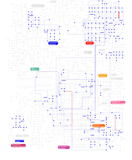
Click the image to view the interactive version of the map in iPath% proteins involved KEGG pathway ID Description 63.24  map00230
Purine metabolism 21.08 map02020 Two-component system - General 8.39 map04914 Progesterone-mediated oocyte maturation 2.74 map00760Nicotinate and nicotinamide metabolism 1.72 map00240Pyrimidine metabolism 0.39 map00260Glycine, serine and threonine metabolism 0.39 map00970 Aminoacyl-tRNA biosynthesis 0.24 map00530Aminosugars metabolism 0.24 map00740Riboflavin metabolism 0.24 map00730Thiamine metabolism 0.24 map00051Fructose and mannose metabolism 0.16 map00791Atrazine degradation 0.08 map00350Tyrosine metabolism 0.08 map00251Glutamate metabolism 0.08 map00903 Limonene and pinene degradation 0.08 map00650Butanoate metabolism 0.08 map00620Pyruvate metabolism 0.08 map006241- and 2-Methylnaphthalene degradation 0.08 map00120Bile acid biosynthesis 0.08 map00471D-Glutamine and D-glutamate metabolism 0.08 map00330Arginine and proline metabolism 0.08 map00460Cyanoamino acid metabolism 0.08 map00632Benzoate degradation via CoA ligation 0.08 map00362Benzoate degradation via hydroxylation This information is based on mapping of SMART genomic protein database to KEGG orthologous groups. Percentage points are related to the number of proteins with HDc domain which could be assigned to a KEGG orthologous group, and not all proteins containing HDc domain. Please note that proteins can be included in multiple pathways, ie. the numbers above will not always add up to 100%.
- Structure (3D structures containing this domain)
3D Structures of HDc domains in PDB
PDB code Main view Title 1f0j 
CATALYTIC DOMAIN OF HUMAN PHOSPHODIESTERASE 4B2B 1mkd 
crystal structure of PDE4D catalytic domain and zardaverine complex 1oyn 
Crystal structure of PDE4D2 in complex with (R,S)-rolipram 1ptw 
The Crystal Structure of AMP-Bound PDE4 Suggests a Mechanism for Phosphodiesterase Catalysis 1q9m 
Three dimensional structures of PDE4D in complex with roliprams and implication on inhibitor selectivity 1rkp 
Crystal structure of PDE5A1-IBMX 1ro6 
Crystal structure of PDE4B2B complexed with Rolipram (R & S) 1ro9 
CRYSTAL STRUCTURES OF THE CATALYTIC DOMAIN OF PHOSPHODIESTERASE 4B2B COMPLEXED WITH 8-Br-AMP 1ror 
CRYSTAL STRUCTURES OF THE CATALYTIC DOMAIN OF PHOSPHODIESTERASE 4B2B COMPLEXED WITH AMP 1so2 
CATALYTIC DOMAIN OF HUMAN PHOSPHODIESTERASE 3B In COMPLEX WITH A DIHYDROPYRIDAZINE INHIBITOR 1soj 
CATALYTIC DOMAIN OF HUMAN PHOSPHODIESTERASE 3B IN COMPLEX WITH IBMX 1t9r 
Catalytic Domain Of Human Phosphodiesterase 5A 1t9s 
Catalytic Domain Of Human Phosphodiesterase 5A in Complex with GMP 1taz 
Catalytic Domain Of Human Phosphodiesterase 1B 1tb5 
Catalytic Domain Of Human Phosphodiesterase 4B In Complex With AMP 1tb7 
Catalytic Domain Of Human Phosphodiesterase 4D In Complex With AMP 1tbb 
Catalytic Domain Of Human Phosphodiesterase 4D In Complex With Rolipram 1tbf 
Catalytic Domain Of Human Phosphodiesterase 5A in Complex with Sildenafil 1udt 
Crystal structure of Human Phosphodiesterase 5 complexed with Sildenafil(Viagra) 1udu 
Crystal structure of Human Phosphodiesterase 5 complexed with tadalafil(Cialis) 1uho 
Crystal structure of Human Phosphodiesterase 5 complexed with Vardenafil(Levitra) 1vj7 
Crystal structure of the bifunctional catalytic fragment of RelSeq, the RelA/SpoT homolog from Streptococcus equisimilis. 1xlx 
Catalytic Domain Of Human Phosphodiesterase 4B In Complex With Cilomilast 1xlz 
Catalytic Domain Of Human Phosphodiesterase 4B In Complex With Filaminast 1xm4 
Catalytic Domain Of Human Phosphodiesterase 4B In Complex With Piclamilast 1xm6 
Catalytic Domain Of Human Phosphodiesterase 4B In Complex With (R)-Mesopram 1xmu 
Catalytic Domain Of Human Phosphodiesterase 4B In Complex With Roflumilast 1xmy 
Catalytic Domain Of Human Phosphodiesterase 4B In Complex With (R)-Rolipram 1xn0 
Catalytic Domain Of Human Phosphodiesterase 4B In Complex With (R,S)-Rolipram 1xom 
Catalytic Domain Of Human Phosphodiesterase 4D In Complex With Cilomilast 1xon 
Catalytic Domain Of Human Phosphodiesterase 4D In Complex With Piclamilast 1xoq 
Catalytic Domain Of Human Phosphodiesterase 4D In Complex With Roflumilast 1xor 
Catalytic Domain Of Human Phosphodiesterase 4D In Complex With Zardaverine 1xos 
Catalytic Domain Of Human Phosphodiesterase 4B In Complex With Sildenafil 1xot 
Catalytic Domain Of Human Phosphodiesterase 4B In Complex With Vardenafil 1xoz 
Catalytic Domain Of Human Phosphodiesterase 5A In Complex With Tadalafil 1xp0 
Catalytic Domain Of Human Phosphodiesterase 5A In Complex With Vardenafil 1xx7 
Conserved hypothetical protein from Pyrococcus furiosus Pfu-403030-001 1y2b 
Catalytic Domain Of Human Phosphodiesterase 4D In Complex With 3,5-dimethyl-1H-pyrazole-4-carboxylic acid ethyl ester 1y2c 
Catalytic Domain Of Human Phosphodiesterase 4D In Complex With 3,5-dimethyl-1-phenyl-1H-pyrazole-4-carboxylic acid ethyl ester 1y2d 
Catalytic Domain Of Human Phosphodiesterase 4D In Complex With 1-(4-methoxy-phenyl)-3,5-dimethyl-1H-pyrazole-4-carboxylic acid ethyl ester 1y2e 
Catalytic Domain Of Human Phosphodiesterase 4D In Complex With 1-(4-amino-phenyl)-3,5-dimethyl-1H-pyrazole-4-carboxylic acid ethyl ester 1y2h 
Catalytic Domain Of Human Phosphodiesterase 4B In Complex With 1-(2-chloro-phenyl)-3,5-dimethyl-1H-pyrazole-4-carboxylic acid ethyl ester 1y2j 
Catalytic Domain Of Human Phosphodiesterase 4B In Complex With 3,5-dimethyl-1-(3-nitro-phenyl)-1H-pyrazole-4-carboxylic acid ethyl ester 1y2k 
Catalytic Domain Of Human Phosphodiesterase 4D In Complex With 3,5-dimethyl-1-(3-nitro-phenyl)-1H-pyrazole-4-carboxylic acid ethyl ester 1ynb 
crystal structure of genomics APC5600 1yoy 
Predicted coding region AF1432 from Archaeoglobus Fulgidus 1z1l 
The Crystal Structure of the Phosphodiesterase 2A Catalytic Domain 1zkl 
Multiple Determinants for Inhibitor Selectivity of Cyclic Nucleotide Phosphodiesterases 1zkn 
Structure of PDE4D2-IBMX 2chm 
Crystal structure of N2 substituted pyrazolo pyrimidinones - a flipped binding mode in PDE5 2cqz 
Crystal Structure of PH0347 protein from Pyrococcus horikoshii OT3 2dqb 
Crystal structure of dNTP triphosphohydrolase from Thermus thermophilus HB8, which is homologous to dGTP triphosphohydrolase 2fm0 
Crystal structure of PDE4D in complex with L-869298 2fm5 
Crystal structure of PDE4D2 in complex with inhibitor L-869299 2h40 
Crystal structure of the catalytic domain of unliganded PDE5 2h42 
Crystal structure of PDE5 in complex with sildenafil 2h44 
Crystal structure of PDE5A1 in complex with icarisid II 2hd1 
Crystal structure of PDE9 in complex with IBMX 2hek 
Crystal structure of O67745, a hypothetical protein from Aquifex aeolicus at 2.0 A resolution. 2o08 
CRYSTAL STRUCTURE OF A PUTATIVE HD SUPERFAMILY HYDROLASE (BH1327) FROM BACILLUS HALODURANS AT 1.90 A RESOLUTION 2o6i 
Structure of an Enterococcus Faecalis HD Domain Phosphohydrolase 2o8h 
Crystal structure of the catalytic domain of rat phosphodiesterase 10A 2ogi 
Crystal structure of a putative metal dependent phosphohydrolase (sag1661) from streptococcus agalactiae serogroup v at 1.85 A resolution 2oun 
crystal structure of PDE10A2 in complex with AMP 2oup 
crystal structure of PDE10A 2ouq 
crystal structure of PDE10A2 in complex with GMP 2our 
crystal structure of PDE10A2 mutant D674A in complex with cAMP 2ous 
crystal structure of PDE10A2 mutant D674A 2ouu 
crystal structure of PDE10A2 mutant D674A in complex with cGMP 2ouv 
crystal structure of pde10a2 mutant of D564N 2ouy 
crystal structure of pde10a2 mutant D564A in complex with cAMP. 2ovv 
Crystal structure of the catalytic domain of rat phosphodiesterase 10A 2ovy 
Crystal structure of the catalytic domain of rat phosphodiesterase 10A 2paq 
Crystal structure of the 5'-deoxynucleotidase YfbR 2par 
Crystal structure of the 5'-deoxynucleotidase YfbR mutant E72A complexed with Co(2+) and TMP 2pau 
Crystal structure of the 5'-deoxynucleotidase YfbR mutant E72A complexed with Co(2+) and dAMP 2pgs 
Crystal structure of a putative deoxyguanosinetriphosphate triphosphohydrolase from Pseudomonas syringae pv. phaseolicola 1448A 2pjq 
Crystal structure of Q88U62_LACPL from Lactobacillus plantarum. Northeast Structural Genomics target LpR71 2pq7 
Crystal structure of predicted HD superfamily hydrolase (104161995) from uncultured Thermotogales bacterium at 1.45 A resolution 2pw3 
Structure of the PDE4D-cAMP complex 2q14 
Crystal structure of Phosphohydrolase (BT4208) from Bacteroides thetaiotaomicron VPI-5482 at 2.20 A resolution 2qgs 
Crystal structure of SE1688 protein from Staphylococcus epidermidis. Northeast Structural Genomics Consortium target SeR89 2qyk 
Crystal structure of PDE4A10 in complex with inhibitor NPV 2qyl 
Crystal structure of PDE4B2B in complex with inhibitor NPV 2qym 
crystal structure of unliganded PDE4C2 2qyn 
Crystal structure of PDE4D2 in complex with inhibitor NPV 2r8q 
Structure of LmjPDEB1 in complex with IBMX 2wey 
Human PDE-papaverine complex obtained by ligand soaking of cross- linked protein crystals 2y0j 
Triazoloquinazolines as a novel class of phosphodiesterase 10A (PDE10A) inhibitors, part 2, Lead-optimisation. 2yy2 
Crystal structure of the human Phosphodiesterase 9A catalytic domain complexed with IBMX 3b2r 
Crystal Structure of PDE5A1 catalytic domain in complex with Vardenafil 3b57 
Crystal structure of the Lin1889 protein (Q92AN1) from Listeria innocua. Northeast Structural Consortium target LkR65 3bg2 
Crystal structure of deoxyguanosinetriphosphate triphosphohydrolase from Flavobacterium sp. MED217 3bjc 
Crystal structure of the PDE5A catalytic domain in complex with a novel inhibitor 3ccg 
Crystal structure of predicted HD superfamily hydrolase involved in NAD metabolism (NP_347894.1) from Clostridium acetobutylicum at 1.50 A resolution 3d3p 
Crystal structure of PDE4B catalytic domain in complex with a pyrazolopyridine inhibitor 3djb 
Crystal structure of a HD-superfamily hydrolase (BT9727_1981) from Bacillus thuringiensis, Northeast Structural Genomics Consortium Target BuR114 3dto 
Crystal structure of the metal-dependent HD domain-containing hydrolase BH2835 from Bacillus halodurans, Northeast Structural Genomics Consortium Target BhR130. 3dy8 
Human Phosphodiesterase 9 in complex with product 5'-GMP (E+P complex) 3dyl 
human phosphdiesterase 9 substrate complex (ES complex) 3dyn 
human phosphodiestrase 9 in complex with cGMP (Zn inhibited) 3dyq 
human phosphodiestrase 9 (inhibited by omitting divalent cation) in complex with cGMP 3dys 
human phosphodiestrase-5'GMP complex (EP), produced by soaking with 20mM cGMP+20 mM MnCl2+20 mM MgCl2 for 2 hours, and flash-cooled to liquid nitrogen temperature when substrate was still abudant. 3ecm 
Crystal structure of the unliganded PDE8A catalytic domain 3ecn 
Crystal structure of PDE8A catalytic domain in complex with IBMX 3frg 
Catalytic Domain of Human Phosphodiesterase 4B2B in Complex with a Quinoline Inhibitor 3g3n 
PDE7A catalytic domain in complex with 3-(2,6-difluorophenyl)-2-(methylthio)quinazolin-4(3H)-one 3g45 
Crystal structure of human phosphodiesterase 4b with regulatory domain and d155988 3g4g 
Crystal structure of human phosphodiesterase 4d with regulatory domain and d155871 3g4i 
Crystal structure of human phosphodiesterase 4d with d155871 3g4k 
Crystal structure of human phosphodiesterase 4d with rolipram 3g4l 
Crystal structure of human phosphodiesterase 4d with roflumilast 3g58 
Crystal structure of human phosphodiesterase 4d with d155988/pmnpq 3gw7 
Crystal structure of a metal-dependent phosphohydrolase with conserved HD domain (yedJ) from Escherichia coli in complex with nickel ions. Northeast Structural Genomics Consortium Target ER63 3gwt 
Catalytic domain of human phosphodiesterase 4B2B in complex with a quinoline inhibitor 3hc1 
Crystal structure of HDOD domain protein with unknown function (NP_953345.1) from GEOBACTER SULFURREDUCENS at 1.90 A resolution 3hc8 
Investigation of Aminopyridiopyrazinones as PDE5 Inhibitors: Evaluation of Modifications to the Central Ring System. 3hdz 
Identification, Synthesis, and SAR of Amino Substituted Pyrido[3,2b]pryaziones as Potent and Selective PDE5 Inhibitors 3hmv 
Catalytic domain of human phosphodiesterase 4B2B in complex with a tetrahydrobenzothiophene inhibitor 3hqw 
Discovery of novel inhibitors of PDE10A 3hqy 
Discovery of novel inhibitors of PDE10A 3hqz 
Discovery of novel inhibitors of PDE10A 3hr1 
Discovery of novel inhibitors of PDE10A 3i8v 
Crystal structure of human PDE4a with 4-(3-butoxy-4-methoxyphenyl)methyl-2-imidazolidone 3iad 
Crystal structure of human phosphodiesterase 4D with bound allosteric modulator 3iak 
Crystal structure of human phosphodiesterase 4d (PDE4d) with papaverine. 3ibj 
X-ray structure of PDE2A 3irh 
Structure of an Enterococcus Faecalis HD-domain protein complexed with dGTP and dATP 3itm 
Catalytic domain of hPDE2A 3itu 
hPDE2A catalytic domain complexed with IBMX 3jab 
3JAB 3jbq 
3JBQ 3jsi 
Human phosphodiesterase 9 in complex with inhibitor 3jsw 
Human PDE9 in complex with selective inhibitor 3jwq 
Crystal structure of chimeric PDE5/PDE6 catalytic domain complexed with sildenafil 3jwr 
Crystal structure of chimeric PDE5/PDE6 catalytic domain complexed with 3-isobutyl-1-methylxanthine (IBMX) and PDE6 gamma-subunit inhibitory peptide 70-87. 3k3e 
Crystal structure of the PDE9A catalytic domain in complex with (R)-BAY73-6691 3k3h 
Crystal structure of the PDE9A catalytic domain in complex with (S)-BAY73-6691 3k4s 
The structure of the catalytic domain of human PDE4d with 4-(3-Butoxy-4-methoxyphenyl)methyl-2-imidazolidone 3kh1 
Crystal structure of Predicted metal-dependent phosphohydrolase (ZP_00055740.2) from Magnetospirillum magnetotacticum MS-1 at 1.37 A resolution 3kkt 
Crystal structure of human PDE4b with 5-[3-[(1S,2S,4R)-Bicyclo[2.2.1]hept-2-yloxy]-4-methoxyp henyl]tetrahydro-2(1H)-pyrimidinone reveals ordering of the C-terminal helix residues 502-509. 3lxg 
Crystal structure of rat phosphodiesterase 10A in complex with ligand WEB-3 3ly2 
Catalytic Domain of Human Phosphodiesterase 4B in Complex with A Coumarin-Based Inhibitor 3mzo 
Crystal structure of a HD-domain phosphohydrolase (lin2634) from LISTERIA INNOCUA at 1.98 A resolution 3n3z 
Crystal structure of PDE9A (E406A) mutant in complex with IBMX 3nqw 
A metazoan ortholog of SpoT hydrolyzes ppGpp and plays a role in starvation responses 3nr1 
A metazoan ortholog of SpoT hydrolyzes ppGpp and plays a role in starvation responses 3o0j 
PDE4B In complex with ligand an2898 3o56 
Catalytic domain of human phosphodiesterase 4b2b in complex with a 5-heterocycle pyrazolopyridine inhibitor 3o57 
Catalytic domain of human phosphodiesterase 4b2b in complex with a 5-heterocycle pyrazolopyridine inhibitor 3qi3 
Crystal structure of PDE9A(Q453E) in complex with inhibitor BAY73-6691 3qi4 
Crystal structure of PDE9A(Q453E) in complex with IBMX 3qpn 
Structure of PDE10-inhibitor complex 3qpo 
Structure of PDE10-inhibitor complex 3qpp 
Structure of PDE10-inhibitor complex 3shy 
Crystal structure of the PDE5A1 catalytic domain in complex with novel inhibitors 3shz 
Crystal structure of the PDE5A1 catalytic domain in complex with novel inhibitors 3sie 
Crystal structure of the PDE5A1 catalytic domain in complex with novel inhibitors 3sk9 
Crystal structure of the Thermus thermophilus cas3 HD domain 3skd 
Crystal structure of the Thermus thermophilus cas3 HD domain in the presence of Ni2+ 3sl3 
Crystal structure of the apo form of the catalytic domain of PDE4D2 3sl4 
Crystal structure of the catalytic domain of PDE4D2 with compound 10D 3sl5 
Crystal structure of the catalytic domain of PDE4D2 complexed with compound 10d 3sl6 
Crystal structure of the catalytic domain of PDE4D2 with compound 12c 3sl8 
Crystal structure of the catalytic domain of PDE4D2 with compound 10o 3sn7 
Highly Potent, Selective, and Orally Active Phosphodiestarase 10A Inhibitors 3sni 
Highly Potent, Selective, and Orally Active Phosphodiestarase 10A Inhibitors 3snl 
Highly Potent, Selective, and Orally Active Phosphodiestarase 10A Inhibitors 3tge 
A novel series of potent and selective PDE5 inhibitor1 3tgg 
A novel series of potent and selective PDE5 inhibitor2 3tm8 
Bd1817, a HDG"Y"P protein from Bdellovibrio bacteriovorus 3tmb 
Bd1817, a HDG"Y"P protein from Bdellovibrio bacteriovorus 3tmc 
Bd1817, a HDG"Y"P protein from Bdellovibrio bacteriovorus 3tmd 
Bd1817, a HDG"Y"P protein from Bdellovibrio bacteriovorus 3tvx 
The structure of PDE4A with pentoxifylline at 2.84A resolution 3u1n 
Structure of the catalytic core of human SAMHD1 3ui7 
Discovery of orally active pyrazoloquinoline as a potent PDE10 inhibitor for the management of schizophrenia 3uuo 
The discovery of potent, selectivity, and orally bioavailable pyrozoloquinolines as PDE10 inhibitors for the treatment of Schizophrenia 3v93 
unliganded structure of TcrPDEC1 catalytic domain 3v94 
TcrPDEC1 catalytic domain in complex with inhibitor wyq16 3v9b 
Crystal structure of the catalytic domain of PDE4D2 with (S)-N-(3-{1-[1-(3-Cyclopropylmethoxy-4-difluoromethoxyphenyl)-2-(1-oxypyridin-4-yl)-ethyl]-1H-pyrazl-3-yl}phenyl)acetamide 3w5e 
Crystal structure of phosphodiesterase 4B in complex with compound 31e 3wd9 
Crystal structure of phosphodiesterase 4B in complex with compound 10f 3wfo 
tRNA processing enzyme (apo form 1) 3wfp 
tRNA processing enzyme (apo form 2) 3wfq 
tRNA processing enzyme complex 1 3wfr 
tRNA processing enzyme complex 2 3wfs 
tRNA processing enzyme complex 3 3wi2 
Crystal structure of PDE10A in complex with inhibitor 3ws8 
3WS8 3ws9 
3WS9 3wyk 
3WYK 3wyl 
3WYL 3wym 
3WYM 4ael 
PDE10A in complex with the inhibitor AZ5 4ajd 
Identification and structural characterization of PDE10 fragment inhibitors 4ajf 
Identification and structural characterization of PDE10 fragment inhibitors 4ajg 
Identification and structural characterization of PDE10 fragment inhibitors 4ajm 
Development of a plate-based optical biosensor methodology to identify PDE10 fragment inhibitors 4bbx 
Discovery of a potent, selective and orally active PDE10A inhibitor for the treatment of schizophrenia 4bzb 
Crystal structure of the tetrameric dGTP-bound SAMHD1 mutant catalytic core 4bzc 
Crystal structure of the tetrameric dGTP-bound wild type SAMHD1 catalytic core 4c1i 
4C1I 4d08 
4D08 4d09 
4D09 4ddl 
PDE10a Crystal Structure Complexed with Novel Inhibitor 4dff 
The SAR development of dihydroimidazoisoquinoline derivatives as phosphodiesterase 10A inhibitors for the treatment of schizophrenia 4dmb 
X-ray structure of human hepatitus C virus NS5A-transactivated protein 2 at the resolution 1.9A, Northeast Structural Genomics Consortium (NESG) Target HR6723 4e90 
Human phosphodiesterase 9 in complex with inhibitors 4fcb 
Potent and Selective Phosphodiesterase 10A Inhibitors 4fcd 
Potent and Selective Phosphodiesterase 10A Inhibitors 4g2j 
Human pde9 in complex with selective compound 4g2l 
Human PDE9 in complex with selective compound 4g2w 
Crystal structure of PDE5A in complex with its inhibitor 4g2y 
Crystal structure of PDE5A complexed with its inhibitor 4gh6 
Crystal structure of the PDE9A catalytic domain in complex with inhibitor 28 4heu 
Crystal Structure of PDE10A with a biaryl ether inhibitor ((1-(3-(4-((1H-benzo[d]imidazol-2-yl)amino)phenoxy)pyridin-2-yl)piperidin-4-yl)methanol) 4hf4 
Crystal Structure of PDE10A with a biaryl ether inhibitor (1-(1-(3-(4-(benzo[d]thiazol-2-ylamino)phenoxy)pyrazin-2-yl)piperidin-4-yl)ethanol) 4htx 
Crystal structure of PDE2 catalytic domain in complex with BAY60-7550 4htz 
Crystal structure of PDE2 catalytic domain in space group P1 4i15 
Crystal structure of TbrPDEB1 4i9z 
Crystal structure of the PDE5A1 catalytic domain in complex with novel inhibitors 4ia0 
Crystal structure of the PDE5A1 catalytic domain in complex with novel inhibitors 4jib 
Crystal structure of of PDE2-inhibitor complex 4kp6 
Crystal structure of human phosphodiesterase 4B (PDE4B) in complex with a [1,3,5]triazine derivative 4l1j 
Three dimensional structure of mutant D143A of human HD domain-containing protein 2, Northeast Structural Genomics Consortium (NESG) Target HR6723 4l7e 
Three dimensional structure of mutant D78A of human HD domain-containing protein 2, Genomics Consortium (NESG) Target HR6723 4l7w 
Crystal structure mutant H77A of human HD domain-containing protein 2, Genomics Consortium (NESG) Target HR6723 4lkq 
Crystal structure of PDE10A2 with fragment ZT017 4llj 
Crystal structure of PDE10A2 with fragment ZT214 4llk 
Crystal structure of PDE10A2 with fragment ZT217 4llp 
Crystal structure of PDE10A2 with fragment ZT401 4llx 
Crystal structure of PDE10A2 with fragment ZT434 4lm0 
Crystal structure of PDE10A2 with fragment ZT448 4lm1 
Crystal structure of PDE10A2 with fragment ZT450 4lm2 
Crystal structure of PDE10A2 with fragment ZT462 4lm3 
Crystal structure of PDE10A2 with fragment ZT464 4lm4 
Crystal structure of PDE10A2 with fragment ZT902 4lrl 
Structure of an Enterococcus Faecalis HD-domain protein complexed with dGTP and dTTP 4mcw 
Metallo-enzyme from P. marina 4md6 
4MD6 4mdz 
Metallo-enzyme from P. marina 4me4 
Metallo-enzyme from P. marina 4mlm 
Crystal Structure of PhnZ from uncultured bacterium HF130_AEPn_1 4mln 
Crystal of PhnZ bound to (R)-2-amino-1-hydroxyethylphosphonic acid 4mrw 
Crystal structure of PDE10A2 with fragment ZT0120 (7-chloroquinolin-4-ol) 4mrz 
Crystal structure of PDE10A2 with fragment ZT0429 (4-methyl-3-nitropyridin-2-amine) 4ms0 
Crystal structure of PDE10A2 with fragment ZT0443 (6-chloropyrimidine-2,4-diamine) 4msa 
Crystal structure of PDE10A2 with fragment ZT0449 (5-nitro-1H-benzimidazole) 4msc 
Crystal structure of PDE10A2 with fragment ZT1595 (2-[(quinolin-7-yloxy)methyl]quinoline) 4mse 
Crystal structure of PDE10A2 with fragment ZT1597 (2-({[(2S)-2-methyl-2,3-dihydro-1,3-benzothiazol-5-yl]oxy}methyl)quinoline) 4msh 
Crystal Structure of PDE10A2 with fragment ZT0143 ((2S)-4-chloro-2,3-dihydro-1,3-benzothiazol-2-amine) 4msn 
Crystal structure of PDE10A2 with fragment ZT0451 (8-nitroquinoline) 4muw 
Crystal Structure of PDE10A with Novel Keto-Benzimidazole Inhibitor 4mvh 
Crystal Structure of PDE10A with Novel Keto-Benzimidazole Inhibitor 4myq 
Selective Inhibition of the Catalytic Domain Of Human Phosphodiesterase 4B With A-33 4mz7 
Structural insight into dGTP-dependent activation of tetrameric SAMHD1 deoxynucleoside triphosphate triphosphohydrolase 4n6w 
X-Ray Crystal Structure of Citrate-bound PhnZ 4n71 
X-Ray Crystal Structure of 2-amino-1-hydroxyethylphosphonate-bound PhnZ 4npv 
4NPV 4npw 
4NPW 4nw7 
4NW7 4oew 
4OEW 4oex 
4OEX 4ogb 
4OGB 4p0n 
4P0N 4p1r 
4P1R 4phl 
4PHL 4phw 
4PHW 4pm0 
4PM0 4q7h 
4Q7H 4qfx 
4QFX 4qfy 
4QFY 4qfz 
4QFZ 4qg0 
4QG0 4qg1 
4QG1 4qg2 
4QG2 4qg4 
4QG4 4qge 
4QGE 4qqw 
4QQW 4qqx 
4QQX 4qqy 
4QQY 4qqz 
4QQZ 4r8z 
4R8Z 4rxo 
4RXO 4rxp 
4RXP 4rxq 
4RXQ 4rxr 
4RXR 4rxs 
4RXS 4s1b 
4S1B 4s1c 
4S1C 4tnp 
4TNP 4tnq 
4TNQ 4tnr 
4TNR 4tnx 
4TNX 4tny 
4TNY 4tnz 
4TNZ 4to0 
4TO0 4to1 
4TO1 4to2 
4TO2 4to3 
4TO3 4to4 
4TO4 4to5 
4TO5 4to6 
4TO6 4tpm 
4TPM 4tpp 
4TPP 4w1o 
4W1O 4wcu 
4WCU 4wn1 
4WN1 4wzi 
4WZI 4x0f 
4X0F 4x9e 
4X9E 4xds 
4XDS 4xy2 
4XY2 4y2b 
4Y2B 4y86 
4Y86 4y87 
4Y87 4y8c 
4Y8C 4yf1 
4YF1 4yqh 
4YQH 4ys7 
4YS7 4zo5 
4ZO5 4zwe 
4ZWE 4zwg 
4ZWG 5ao0 
5AO0 5ao1 
5AO1 5ao2 
5AO2 5ao3 
5AO3 5ao4 
5AO4 5axp 
5AXP 5axq 
5AXQ 5b25 
5B25 5b4k 
5B4K 5b4l 
5B4L 5b7i 
5B7I 5c1w 
5C1W 5c28 
5C28 5c29 
5C29 5c2a 
5C2A 5c2e 
5C2E 5c2h 
5C2H 5dh4 
5DH4 5dh5 
5DH5 5dqv 
5DQV 5dqw 
5DQW 5ede 
5EDE 5edg 
5EDG 5edh 
5EDH 5edi 
5EDI 5gqh 
5GQH 5i2r 
5I2R 5ihy 
5IHY 5k9r 
5K9R 5tk6 
5TK6 5tk7 
5TK7 5tk8 
5TK8 5tk9 
5TK9 5tka 
5TKA - Links (links to other resources describing this domain)
-
INTERPRO IPR003607