Chorismate mutase, catalyses the conversion of chorismate to prephenate in the pathway of tyrosine and phenylalanine biosynthesis. This enzyme is negatively regulated by tyrosine, tryptophan and phenylalanine (PUBMED:9642265), (PUBMED:9497350).
Chorismate mutase (CM) is a regulatory enzyme ( EC 5.4.99.5 ) required for biosynthesis of the aromatic amino acids phenylalanine and tyrosine. CM catalyzes the Claisen rearrangement of chorismate to prephenate, which can subsequently be converted to precursors of either L-Phe or L-Tyr. In bifunctional enzymes the CM domain can be fused to a prephenate dehydratase (P-protein for Phe biosynthesis), to a prephenate dehydrogenase (T-protein, for Tyr biosynthesis), or to 3-deoxy-D-arabino-heptulosonate 7-phosphate (DAHP) synthase. Besides these prokaryotic bifunctional enzymes, monofunctional CMs occur in prokaryotes as well as in fungi, plants and nematode worms [ (PUBMED:11528003) ]. The sequence of monofunctional chorismate mutase aligns well with the N-terminal part of P-proteins [ (PUBMED:9642265) ].
The type II or AroQ class of CM has an all-helical 3D structure, represented by the CM domain of the bifunctional Escherichia coli P-protein. This type is named after the Enterobacter agglomerans monofunctional CM encoded by the aroQ gene [ (PUBMED:8335631) ]. All CM domains from bifunctional enzymes as well as most monofunctional CMs belong to this class, including archaeal CM.
Eukaryotic CM from plants and fungi form a separate subclass of AroQ, represented by the Baker's yeast allosteric CM. These enzymes show only partial sequence similarity to the prokaryotic CMs due to insertions of regulatory domains, but the helix-bundle topology and catalytic residues are conserved and the 3D structure of the E. coli CM dimer resembles a yeast CM monomer [ (PUBMED:11528003) (PUBMED:9384560) (PUBMED:9665711) ]. The E. coli P-protein CM domain consists of 3 helices and lacks allosteric regulation. The yeast CM has evolved by gene duplication and dimerization and each monomer has 12 helices. Yeast CM is allosterically activated by Trp and inhibited by Tyr [ (PUBMED:9384560) ].
This entry represents the CM type 2 domain, mainly from prokaryotes. It does not include the CM from plants and or Baker's yeast.
Tyrosine and tryptophan act through the same binding site at the dimerinterface of yeast chorismate mutase.
J Biol Chem. 1998; 273: 17012-7
Display abstract
Tyrosine and tryptophan are the regulators of the dimeric yeast chorismatemutase. Biochemical studies reveal two binding sites per molecule for botheffectors, tyrosine or tryptophan. A single binding site is built up byhelix 8 and helices 4 and 5 of two different subunits. The binding siteshave been analyzed in the active enzyme by site directed mutagenesis ofcritical codons of the coding gene, ARO7. Gly-141 and Ser-142, which bothreside on helix 8, are involved in the binding of tyrosine or tryptophanpresumably by interacting specifically with the amino- andcarboxylate-groups of these amino acid effectors. Interaction with Thr-145of helix 8 is required for a strong tyrosine binding to the allostericsite. Replacement of Arg-75, which connects helices 4 and 5 or of Arg-76,which is part of helix 5 by alanine residues, resulted in unregulatedenzymes. These two residues are bonded to the carboxylate group andphenolic hydroxyl group of tyrosine, respectively, but do not interactwith tryptophan by hydrogen bonding in the crystal structures.Phenylalanine, which has low binding affinity slightly activated thechorismate mutase. A T145V mutant chorismate mutase, however, showedincreased activation by phenylalanine. Our results support a mechanism bywhich tyrosine contracts the allosteric site by interacting with itsphenolic hydroxyl group. Tryptophan works in an inverse way by opening theallosteric site through the steric size of its side chain.
Chorismate mutase-prephenate dehydratase from Escherichia coli. Study ofcatalytic and regulatory domains using genetically engineered proteins.
J Biol Chem. 1998; 273: 6248-53
Display abstract
The bifunctional P-protein, which plays a central role in Escherichia coliphenylalanine biosynthesis, contains two catalytic domains (chorismatemutase and prephenate dehydratase activities) as well as one R-domain (forfeedback inhibition by phenylalanine). Six genes coding for P-proteindomains or subdomains were constructed and successfully expressed.Proteins containing residues 1-285 and residues 1-300 retained full mutaseand dehydratase activity, but exhibited no feedback inhibition. Proteinscontaining residues 101-386 and residues 101-300 retained full dehydrataseactivity, but lacked mutase activity. Fluorescence emission spectra andbinding assays indicated that residues 286-386 were crucial forphenylalanine binding. The mutase (residues 1-109), dehydratase (residues101-285), and regulatory (residues 286-386) activities were thus shown toreside in discrete domains of the P-protein. Both the mutase domain andthe native P-protein formed dimers. Deletion of the mutase domaindiminished phenylalanine binding to the regulatory site as well asprephenate binding to the dehydratase domain, both through cooperativeeffects. Besides eliminating feedback inhibition, removal of the R-domaindecreased the affinity of chorismate mutase for chorismate.
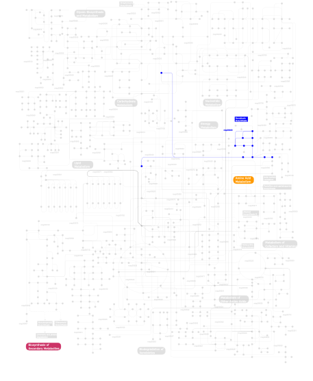
Metabolism (metabolic pathways involving proteins which contain this domain)
Click the image to view the interactive version of the map in iPath
This information is based on mapping of SMART genomic protein database to KEGG orthologous groups. Percentage points are related to the number of proteins with CM_2 domain which could be assigned to a KEGG orthologous group, and not all proteins containing CM_2 domain. Please note that proteins can be included in multiple pathways, ie. the numbers above will not always add up to 100%.
The 2.07 Angstrom crystal structure of Mycobacterium tuberculosis chorismate mutase reveals unexpected gene duplication and suggests a role in host-pathogen interactions
1.95 Angstrom crystal structure of a bifunctional 3-deoxy-7-phosphoheptulonate synthase/chorismate mutase (aroA) from Listeria monocytogenes EGD-e in complex with phosphoenolpyruvate