Asparaginase |
![]() |
|---|
| SMART accession number: | SM00870 |
|---|---|
| Description: | Asparaginase, which is found in various plant, animal and bacterial cells, catalyses the deamination of asparagine to yield aspartic acid and an ammonium ion, resulting in a depletion of free circulatory asparagine in plasma (PUBMED:3026924). The enzyme is effective in the treatment of human malignant lymphomas, which have a diminished capacity to produce asparagine synthetase: in order to survive, such cells absorb asparagine from blood plasma (PUBMED:2407723), (PUBMED:3379033) - if Asn levels have been depleted by injection of asparaginase, the lymphoma cells die. |
| Family alignment: |
There are 697 Asparaginase domains in 697 proteins in SMART's nrdb database.
Click on the following links for more information.
- Evolution (species in which this domain is found)
-
Click on
to expand nodes. To display all proteins with a Asparaginase domain in a specific node, click on it.This tree shows only several representative species. The complete taxonomic breakdown of all proteins with Asparaginase domain is also avaliable.
Useful shortcuts: Expand all nodes or Collapse tree
Go to specific node: Anopheles gambiae, Caenorhabditis elegans, Drosophila melanogaster, Homo sapiens, Mus musculus, Rattus norvegicus, Saccharomyces cerevisiae, Takifugu rubripes - Cellular role (predicted cellular role)
-
Cellular role: metabolism
- Literature (relevant references for this domain)
-
Primary literature is listed below; Automatically-derived, secondary literature is also avaliable.
- Jennings MP, Beacham IR
- Analysis of the Escherichia coli gene encoding L-asparaginase II, ansB,and its regulation by cyclic AMP receptor and FNR proteins.
- J Bacteriol. 1990; 172: 1491-8
- Display abstract
Escherichia coli contains two L-asparaginase isozymes: L-asparaginase I, alow-affinity enzyme located in the cytoplasm, and L-asparaginase II, ahigh-affinity secreted enzyme. A molecular genetic analysis of the gene(ansA) encoding the former enzyme has previously been reported. We nowpresent a molecular study of the gene, ansB, encoding L-asparaginase II.This gene was isolated by using oligonucleotide probes, whose sequenceswere based on the previously determined amino acid sequence. Thenucleotide sequence of ansB, including 5'- and 3'-untranslated regions,was determined. The amino acid sequence of L-asparaginase II, deduced fromthis nucleotide sequence, contains differences at 11 positions whencompared with the previously determined amino acid sequence. The deducedamino acid sequence also reveals a typical secretory signal peptide of 22residues. A single region of sequence similarity is observed when ansA andansB are compared. The transcriptional start site in ansB was determined,allowing the identification of the promoter region. The regulation of ansBwas studied by using ansB'-'lacZ fusions, together with a deletionanalysis of the 5' region upstream of the promoter. Regulation by cyclicAMP receptor protein and anaerobiosis (FNR protein) was confirmed, and thepresence of nucleotide sequence motifs, with homology to cyclic AMPreceptor protein and FNR protein-binding sites, investigated.
- Tanaka S et al.
- Structures of amidohydrolases. Amino acid sequence of aglutaminase-asparaginase from Acinetobacter glutaminasificans andpreliminary crystallographic data for an asparaginase from Erwiniachrysanthemi.
- J Biol Chem. 1988; 263: 8583-91
- Display abstract
The complete amino acid sequence of a glutaminase-asparaginase fromAcinetobacter glutaminasificans, for which a preliminary tertiarystructure is available from crystallographic analysis, has been determinedby automated Edman degradation of fragments produced by chemical andproteolytic cleavages. The protein consists of 331 amino acid residues andhas a molecular weight of 35,500. The pattern of hydrophilic andhydrophobic regions is typical of a globular protein. A new crystal formof an Erwinia chrysanthemi 1125 asparaginase is reported. The space groupis monoclinic C2, with unit cell parameters of: a = 107.8, b = 91.7, c =129.2 A and beta = 91.7 degrees. A Vm of 2.25 A3/dalton was calculated forone tetramer of 35,100-dalton subunits per asymmetric unit. X-rayintensity data have been obtained to 2.2 A resolution. The point groupsymmetry of the Er. chrysanthemi tetramer is 222 from self-rotationfunction calculations. The relative orientations of an A.glutaminasificans glutaminase-asparaginase model and the Er. chrysanthemiasparaginase tetramer have been determined with the cross-rotationfunction, and translation function calculations have revealed a plausiblelocation for the asparaginase tetramer in the crystal.
- Minton NP, Bullman HM, Scawen MD, Atkinson T, Gilbert HJ
- Nucleotide sequence of the Erwinia chrysanthemi NCPPB 1066 L-asparaginasegene.
- Gene. 1986; 46: 25-35
- Display abstract
The complete nucleotide sequence of the Erwinia chrysanthemi NCPPB 1066gene coding for the chemotherapeutic enzyme L-asparaginase has beendetermined. The structural gene consists of an open reading framecommencing with an ATG start codon of 1044 bp followed by a TGA stopcodon. Confirmation of the nucleotide sequence was obtained by comparingthe predicted amino acid (aa) sequence with that derived by N-terminal aasequencing of the purified protein. The gene has been shown to code for a21-aa signal peptide at its N terminus which closely resembles the signalpeptides of other secreted proteins. In common with highly expressedEscherichia coli genes, little use is made of modulator codons. Thepredicted aa sequence of the enzyme exhibits 46% identity with thedetermined primary sequence of the E. coli L-asparaginase, although thepredicted secondary structure of both proteins indicates more extensivehomology. Downstream of the TGA stop codon is a G + C-rich region of dyadsymmetry (delta G = -25.4 kcal) characteristic of E. coli Rho-independenttranscription terminators. Upstream of the structural gene there are nosequences which bear a strong resemblance to the consensus -35 and -10regions of E. coli promoters. A sequence is present (CTGGCTCTCCTCTTGAT),however, which exhibits strong homology to the nif promoter consensussequence (CTGGCACN5TTGCA). Upstream of this region is a sequence whichstrongly resembles the consensus sequence for promoter regions which aresubject to catabolite repression.
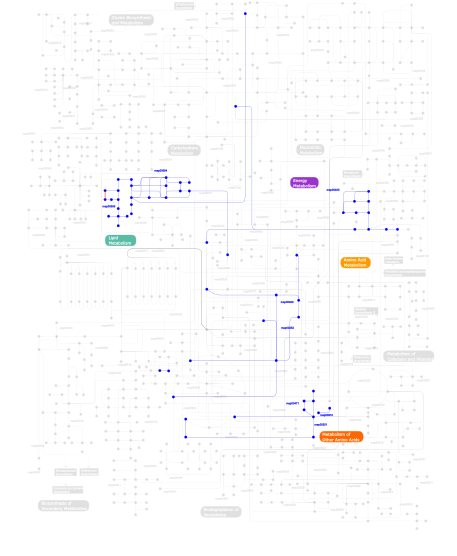
- Metabolism (metabolic pathways involving proteins which contain this domain)
-

Click the image to view the interactive version of the map in iPath% proteins involved KEGG pathway ID Description 31.57
map00910Nitrogen metabolism 31.57
map00252Alanine and aspartate metabolism 29.87
map00460Cyanoamino acid metabolism 4.66
map00251Glutamate metabolism 1.69
map00471D-Glutamine and D-glutamate metabolism 0.21
map00565Ether lipid metabolism 0.21
map00564Glycerophospholipid metabolism 0.11
map00400Phenylalanine, tyrosine and tryptophan biosynthesis 0.11 map02020 Two-component system - General This information is based on mapping of SMART genomic protein database to KEGG orthologous groups. Percentage points are related to the number of proteins with Asparaginase domain which could be assigned to a KEGG orthologous group, and not all proteins containing Asparaginase domain. Please note that proteins can be included in multiple pathways, ie. the numbers above will not always add up to 100%.
- Structure (3D structures containing this domain)
3D Structures of Asparaginase domains in PDB
PDB code Main view Title 1agx 
Refined crystal structure of acinetobacter glutaminasificans glutaminase-asparaginase 1djo 
Crystal structure of pseudomonas 7a glutaminase- asparaginase with the inhibitor donv covalently bound in the active site 1djp 
Crystal structure of pseudomonas 7a glutaminase- asparaginase with the inhibitor don covalently bound in the active site 1hfj 
Asparaginase from erwinia chrysanthemi, hexagonal form with sulfate 1hfk 
Asparaginase from erwinia chrysanthemi, hexagonal form with weak sulfate 1hfw 
X-ray structure of the complex between erwinia chrysanthemi l-asparaginase and l-glutamate 1hg0 
X-ray structure of the complex between erwinia chrysanthemi l-asparaginase and succinic acid 1hg1 
X-ray structure of the complex between erwinia chrysanthemi l-asparaginase and d-aspartate 1ho3 
Crystal structure analysis of e. coli l-asparaginase ii (y25f mutant) 1ihd 
Crystal structure of trigonal form of d90e mutant of escherichia coli asparaginase ii 1jaz 
Crystal structure of monoclinic form of d90e mutant of escherichia coli asparaginase ii 1jja 
Crystal structure of orthorhombic form of d90e mutant of escherichia coli l-asparaginase ii 1jsl 
Crystal structure of erwinia chrysanthemi l-asparaginase complexed with 6-hydroxy-d-norleucine 1jsr 
Crystal structure of erwinia chrysanthemi l-asparaginase complexed with 6-hydroxy-l-norleucine 1nns 
L-asparaginase of e. coli in c2 space group and 1.95 a resolution 1o7j 
Atomic resolution structure of erwinia chrysanthemi l- asparaginase 1wls 
Crystal structure of l-asparaginase i homologue protein from pyrococcus horikoshii 1wnf 
Crystal structure of ph0066 from pyrococcus horikoshii 1wsa 
Structure of l-asparaginase ii precursor 1zcf 
L-asparaginase from erwinia carotovora 1zq1 
Structure of gatde trna-dependent amidotransferase from pyrococcus abyssi 2d6f 
Crystal structure of glu-trna(gln) amidotransferase in the complex with trna(gln) 2gvn 
L-asparaginase from erwinia carotovora in complex with aspartic acid 2him 
Crystal structure and allosteric regulation of the cytoplasmic escherichia coli l-asparaginase i 2hln 
L-asparaginase from erwinia carotovora in complex with glutamic acid 2jk0 
Structural and functional insights into erwinia carotovora l-asparaginase 2ocd 
Crystal structure of l-asparaginase i from vibrio cholerae o1 biovar eltor str. n16961 2p2d 
Crystal structure and allosteric regulation of the cytoplasmic escherichia coli l-asparaginase i 2p2n 
Crystal structure and allosteric regulation of the cytoplasmic escherichia coli l-asparaginase i 2wlt 
2wt4 
3eca 
Crystal structure of escherichia coli l-asparaginase, an enzyme used in cancer therapy 3pga 
Structural characterization of pseudomonas 7a glutaminase- asparaginase 4eca 
Asparaginase from e. coli, mutant t89v with covalently bound aspartate 4pga 
Glutaminase-asparaginase from pseudomonas 7a - Links (links to other resources describing this domain)
-
PFAM Asparaginase


