A family of bacterial and eukaryotic endonucleases EC 3.1.30 share the following characteristics: they act on both DNA and RNA, cleave double-stranded and single-stranded nucleic acids and require a divalent ion such as magnesium for their activity. A histidine has been shown [ (PUBMED:8078761) ] to be essential for the activity of the Serratia marcescens nuclease. This residue is located in a conserved region which also contains an aspartic acid residue that could be implicated in the binding of the divalent ion.
2.1 A structure of Serratia endonuclease suggests a mechanism for binding to double-stranded DNA.
Nat Struct Biol. 1994; 1: 461-8
Display abstract
The crystal structure of Serratia endonuclease has been solved to 2.1 A by multiple isomorphous replacement. This magnesium-dependent enzyme is equally active against single- and double-stranded DNA, as well as RNA, without any apparent base preference. The Serratia endonuclease fold is distinct from that of other nucleases that have been solved by X-ray diffraction. The refined structure consists of a central layer containing six antiparallel beta-strands which is flanked on one side by a helical domain and on the opposite side by one dominant helix and a very long coiled loop. Electrostatic calculations reveal a strongly polarized molecular surface and suggest that a cleft between this long helix and loop, near His 89, may contain the active site of the enzyme.
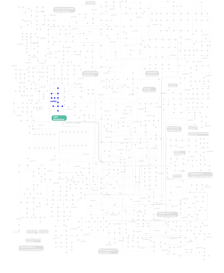
Metabolism (metabolic pathways involving proteins which contain this domain)
Click the image to view the interactive version of the map in iPath
This information is based on mapping of SMART genomic protein database to KEGG orthologous groups. Percentage points are related to the number of proteins with NUC domain which could be assigned to a KEGG orthologous group, and not all proteins containing NUC domain. Please note that proteins can be included in multiple pathways, ie. the numbers above will not always add up to 100%.