TPRTetratricopeptide repeats |
|---|
| SMART accession number: | SM00028 |
|---|---|
| Description: | Repeats present in 4 or more copies in proteins. Contain a minimum of 34 amino acids each and self-associate via a "knobs and holes" mechanism. |
| Interpro abstract (IPR019734): | The tetratrico peptide repeat region (TPR) is a structural motif present in a wide range of proteins [ (PUBMED:7667876) (PUBMED:9482716) (PUBMED:1882418) ]. It mediates protein-protein interactions and the assembly of multiprotein complexes [ (PUBMED:14659697) ]. The TPR motif consists of 3-16 tandem-repeats of 34 amino acids residues, although individual TPR motifs can be dispersed in the protein sequence. Sequence alignment of the TPR domains reveals a consensus sequence defined by a pattern of small and large amino acids. TPR motifs have been identified in various different organisms, ranging from bacteria to humans. Proteins containing TPRs are involved in a variety of biological processes, such as cell cycle regulation, transcriptional control, mitochondrial and peroxisomal protein transport, neurogenesis and protein folding. The X-ray structure of a domain containing three TPRs from protein phosphatase 5 revealed that TPR adopts a helix-turn-helix arrangement, with adjacent TPR motifs packing in a parallel fashion, resulting in a spiral of repeating anti-parallel alpha-helices [ (PUBMED:14659697) ]. The two helices are denoted helix A and helix B. The packing angle between helix A and helix B is ~24 degrees within a single TPR and generates a right-handed superhelical shape. Helix A interacts with helix B and with helix A' of the next TPR. Two protein surfaces are generated: the inner concave surface is contributed to mainly by residue on helices A, and the other surface presents residues from both helices A and B. |
| GO function: | protein binding (GO:0005515) |
| Family alignment: |
There are 1309452 TPR domains in 257843 proteins in SMART's nrdb database.
Click on the following links for more information.
- Evolution (species in which this domain is found)
-
Taxonomic distribution of proteins containing TPR domain.
This tree includes only several representative species. The complete taxonomic breakdown of all proteins with TPR domain is also avaliable.
Click on the protein counts, or double click on taxonomic names to display all proteins containing TPR domain in the selected taxonomic class.
- Cellular role (predicted cellular role)
-
Binding / catalysis: protein-binding
- Literature (relevant references for this domain)
-
Primary literature is listed below; Automatically-derived, secondary literature is also avaliable.
- Das AK, Cohen PW, Barford D
- The structure of the tetratricopeptide repeats of protein phosphatase 5: implications for TPR-mediated protein-protein interactions.
- EMBO J. 1998; 17: 1192-9
- Display abstract
The tetratricopeptide repeat (TPR) is a degenerate 34 amino acid sequence identified in a wide variety of proteins, present in tandem arrays of 3-16 motifs, which form scaffolds to mediate protein-protein interactions and often the assembly of multiprotein complexes. TPR-containing proteins include the anaphase promoting complex (APC) subunits cdc16, cdc23 and cdc27, the NADPH oxidase subunit p67 phox, hsp90-binding immunophilins, transcription factors, the PKR protein kinase inhibitor, and peroxisomal and mitochondrial import proteins. Here, we report the crystal structure of the TPR domain of a protein phosphatase, PP5. Each of the three TPR motifs of this domain consist of a pair of antiparallel alpha-helices of equivalent length. Adjacent TPR motifs are packed together in a parallel arrangement such that a tandem TPR motif structure is composed of a regular series of antiparallel alpha-helices. The uniform angular and spatial arrangement of neighbouring alpha-helices defines a helical structure and creates an amphipathic groove. Multiple-TPR motif proteins would fold into a right-handed super-helical structure with a continuous helical groove suitable for the recognition of target proteins, hence defining a novel mechanism for protein recognition. The spatial arrangement of alpha-helices in the PP5-TPR domain is similar to those within 14-3-3 proteins.
- Lamb JR, Tugendreich S, Hieter P
- Tetratrico peptide repeat interactions: to TPR or not to TPR?
- Trends Biochem Sci. 1995; 20: 257-9
- Goebl M, Yanagida M
- The TPR snap helix: a novel protein repeat motif from mitosis to transcription.
- Trends Biochem Sci. 1991; 16: 173-7
- Display abstract
The recently discovered TPR gene family encodes a diverse group of proteins that function in mitosis, transcription, splicing, protein import and neurogenesis. These multi-domain proteins all contain tandemly arranged repeats of a 34-amino acid motif that are presumed to form helix-turn structures, each with a 'knob' and 'hole', acting as helix-associating domains.
- Sikorski RS, Boguski MS, Goebl M, Hieter P
- A repeating amino acid motif in CDC23 defines a family of proteins and a new relationship among genes required for mitosis and RNA synthesis.
- Cell. 1990; 60: 307-17
- Display abstract
We have identified and characterized a novel, repeating 34 amino acid motif (the TPR motif) that is reiterated several times within the CDC23 gene product of S. cerevisiae. Multiple copies of this motif were discovered in five other proteins, three encoded by cell division cycle genes required to complete mitosis and two involved in RNA synthesis. Quantitative sequence analyses suggest the existence of a common underlying structure in each TPR unit that consists of amphipathic alpha-helical regions punctuated by proline-induced turns. The TPR motif defines a new family of genes and an important structural unit common to several proteins whose functions are required for mitosis and RNA synthesis.
- Disease (disease genes where sequence variants are found in this domain)
-
SwissProt sequences and OMIM curated human diseases associated with missense mutations within the TPR domain.
Protein Disease Peroxisomal targeting signal 1 receptor (P50542) (SMART) OMIM:600414: Adrenoleukodystrophy, neonatal
OMIM:202370:Neutrophil cytosol factor 2 (P19878) (SMART) OMIM:233710: Chronic granulomatous disease due to deficiency of NCF-2 - Metabolism (metabolic pathways involving proteins which contain this domain)
-

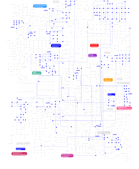
Click the image to view the interactive version of the map in iPath% proteins involved KEGG pathway ID Description 14.61 map04120 Ubiquitin mediated proteolysis 14.38 map04111 Cell cycle - yeast 13.70 map03090 Type II secretion system 13.01 map04110 Cell cycle 13.01 map04914 Progesterone-mediated oocyte maturation 3.65  map00230
Purine metabolism 3.20 map04010 MAPK signaling pathway 2.51 map04020 Calcium signaling pathway 2.28 map01030 Glycan structures - biosynthesis 1 2.28  map00632
Benzoate degradation via CoA ligation 2.28  map00512
O-Glycan biosynthesis 2.05  map00562
Inositol phosphate metabolism 2.05 map02030 Bacterial chemotaxis - General 1.60 map04670 Leukocyte transendothelial migration 1.60  map00051
Fructose and mannose metabolism 1.60  map00561
Glycerolipid metabolism 1.14  map00860
Porphyrin and chlorophyll metabolism 0.68 map00960 Alkaloid biosynthesis II 0.46  map00650
Butanoate metabolism 0.46  map00623
2,4-Dichlorobenzoate degradation 0.46  map00100
Biosynthesis of steroids 0.23  map00350
Tyrosine metabolism 0.23  map00910
Nitrogen metabolism 0.23  map00340
Histidine metabolism 0.23  map00642
Ethylbenzene degradation 0.23 map03060 Protein export 0.23 map03020 RNA polymerase 0.23  map00564
Glycerophospholipid metabolism 0.23 map00903 Limonene and pinene degradation 0.23  map00310
Lysine degradation 0.23  map00624
1- and 2-Methylnaphthalene degradation 0.23  map00360
Phenylalanine metabolism 0.23  map00500
Starch and sucrose metabolism 0.23  map00540
Lipopolysaccharide biosynthesis This information is based on mapping of SMART genomic protein database to KEGG orthologous groups. Percentage points are related to the number of proteins with TPR domain which could be assigned to a KEGG orthologous group, and not all proteins containing TPR domain. Please note that proteins can be included in multiple pathways, ie. the numbers above will not always add up to 100%.
- Structure (3D structures containing this domain)
3D Structures of TPR domains in PDB
PDB code Main view Title 1a17 
TETRATRICOPEPTIDE REPEATS OF PROTEIN PHOSPHATASE 5 1e96 
Structure of the Rac/p67phox complex 1elr 
Crystal structure of the TPR2A domain of HOP in complex with the HSP90 peptide MEEVD 1elw 
Crystal structure of the TPR1 domain of HOP in complex with a HSC70 peptide 1fch 
CRYSTAL STRUCTURE OF THE PTS1 COMPLEXED TO THE TPR REGION OF HUMAN PEX5 1hh8 
crystal structure of the N-terminal region of the phagocyte oxidase factor p67phox at 1.8 Ã… resolution 1ihg 
Bovine Cyclophilin 40, monoclinic form 1iip 
Bovine Cyclophilin 40, Tetragonal Form 1na0 
Design of Stable alpha-Helical Arrays from an Idealized TPR Motif 1p5q 
Crystal Structure of FKBP52 C-terminal Domain 1qz2 
Crystal Structure of FKBP52 C-terminal Domain complex with the C-terminal peptide MEEVD of Hsp90 1w3b 
The superhelical TPR domain of O-linked GlcNAc transferase reveals structural similarities to importin alpha. 1wao 
PP5 structure 1wm5 
Crystal structure of the N-terminal TPR domain (1-203) of p67phox 1xnf 
Crystal structure of E.coli TPR-protein NlpI 2bug 
Solution structure of the TPR domain from Protein phosphatase 5 in complex with Hsp90 derived peptide 2c0l 
TPR DOMAIN OF HUMAN PEX5P IN COMPLEX WITH HUMAN MSCP2 2c0m 
apo form of the TPR domain of the pex5p receptor 2c2l 
Crystal structure of the CHIP U-box E3 ubiquitin ligase 2dba 
The solution structure of the tetratrico peptide repeat of human Smooth muscle cell associated protein-1, isoform 2 2fbn 
Plasmodium falciparum putative FK506-binding protein PFL2275c, C-terminal TPR-containing domain 2fi7 
Crystal Structure of PilF : Functional implication in the type 4 pilus biogenesis in Pseudomonas aeruginosa 2fo7 
Crystal structure of an 8 repeat consensus TPR superhelix (trigonal crystal form) 2gw1 
Crystal Structure of the Yeast Tom70 2ho1 
Functional Characterization of Pseudomonas Aeruginosa pilF 2hyz 
Crystal structure of an 8 repeat consensus TPR superhelix (orthorombic crystal form) 2if4 
Crystal structure of a multi-domain immunophilin from Arabidopsis thaliana 2j9q 
A novel conformation for the TPR domain of pex5p 2jlb 
Xanthomonas campestris putative OGT (XCC0866), complex with UDP- GlcNAc phosphonate analogue 2lni 
Solution NMR Structure of Stress-induced-phosphoprotein 1 STI1 from Homo sapiens, Northeast Structural Genomics Consortium Target HR4403E 2pl2 
Crystal structure of TTC0263: a thermophilic TPR protein in Thermus thermophilus HB27 2q7f 
Crystal structure of YrrB: a TPR protein with an unusual peptide-binding site 2vq2 
Crystal structure of PilW, widely conserved type IV pilus biogenesis factor 2vsn 
Structure and topological arrangement of an O-GlcNAc transferase homolog: insight into molecular control of intracellular glycosylation 2vsy 
Xanthomonas campestris putative OGT (XCC0866), apostructure 2vyi 
Crystal Structure of the TPR domain of Human SGT 2wqh 
Crystal structure of CTPR3Y3 2xev 
Crystal structure of the TPR domain of Xanthomonas campestris ybgF 2xgm 
Substrate and product analogues as human O-GlcNAc transferase inhibitors. 2xgo 
XcOGT in complex with UDP-S-GlcNAc 2xgs 
XcOGT in complex with C-UDP 2xpi 
Crystal structure of APC/C hetero-tetramer Cut9-Hcn1 2y4t 
Crystal structure of the human co-chaperone P58(IPK) 2y4u 
Crystal structure of human P58(IPK) in space group P312 3as4 
MamA AMB-1 C2221 3as5 
MamA AMB-1 P212121 3as8 
MamA MSR-1 P41212 3asd 
MamA R50E mutant 3asf 
MamA MSR-1 C2 3asg 
MamA D159K mutant 2 3ash 
MamA D159K mutant 1 3ceq 
The TPR domain of Human Kinesin Light Chain 2 (hKLC2) 3cv0 
Structure of Peroxisomal Targeting Signal 1 (PTS1) binding domain of Trypanosoma brucei Peroxin 5 (TbPEX5)complexed to T. brucei Phosphoglucoisomerase (PGI) PTS1 peptide 3cvl 
Structure of Peroxisomal Targeting Signal 1 (PTS1) binding domain of Trypanosoma brucei Peroxin 5 (TbPEX5)complexed to T. brucei Phosphofructokinase (PFK) PTS1 peptide 3cvn 
Structure of Peroxisomal Targeting Signal 1 (PTS1) binding domain of Trypanosoma brucei Peroxin 5 (TbPEX5)complexed to T. brucei Glyceraldehyde-3-phosphate dehydrogenase (GAPDH) PTS1 peptide 3cvp 
Structure of Peroxisomal Targeting Signal 1 (PTS1) binding domain of Trypanosoma brucei Peroxin 5 (TbPEX5)complexed to PTS1 peptide (10-SKL) 3cvq 
Structure of Peroxisomal Targeting Signal 1 (PTS1) binding domain of Trypanosoma brucei Peroxin 5 (TbPEX5)complexed to PTS1 peptide (7-SKL) 3edt 
Crystal structure of the mutated S328N hKLC2 TPR domain 3esk 
Structure of HOP TPR2A domain in complex with the non-cognate Hsc70 peptide ligand 3fp2 
Crystal structure of Tom71 complexed with Hsp82 C-terminal fragment 3fp3 
Crystal structure of Tom71 3fp4 
Crystal structure of Tom71 complexed with Ssa1 C-terminal fragment 3fwv 
Crystal Structure of a Redesigned TPR Protein, T-MOD(VMY), in Complex with MEEVF Peptide 3hym 
Insights into Anaphase Promoting Complex TPR subdomain assembly from a CDC26-APC6 structure 3ieg 
Crystal Structure of P58(IPK) TPR Domain at 2.5 A 3jcm 
3JCM 3kd7 
Designed TPR module (CTPR390) in complex with its peptide-ligand (Hsp90 peptide) 3lca 
Structure of Tom71 complexed with Hsp70 Ssa1 C terminal tail indicating conformational plasticity 3nf1 
Crystal structure of the TPR domain of kinesin light chain 1 3pe3 
Structure of human O-GlcNAc transferase and its complex with a peptide substrate 3pe4 
Structure of human O-GlcNAc transferase and its complex with a peptide substrate 3q15 
Crystal Structure of RapH complexed with Spo0F 3q47 
Crystal structure of TPR domain of CHIP complexed with pseudophosphorylated Smad1 peptide 3q49 
Crystal structure of the TPR domain of CHIP complexed with Hsp70-C peptide 3q4a 
Crystal structure of the TPR domain of CHIP complexed with phosphorylated Smad1 peptide 3qky 
Crystal structure of Rhodothermus marinus BamD 3r9a 
Human alanine-glyoxylate aminotransferase in complex with the TPR domain of human PEX5P 3ro2 
Structures of the LGN/NuMA complex 3ro3 
crystal structure of LGN/mInscuteable complex 3sf4 
Crystal structure of the complex between the conserved cell polarity proteins Inscuteable and LGN 3sz7 
Crystal structure of the Sgt2 TPR domain from Aspergillus fumigatus 3tax 
A Neutral Diphosphate Mimic Crosslinks the Active Site of Human O-GlcNAc Transferase 3ulq 
Crystal Structure of the Anti-Activator RapF Complexed with the Response Regulator ComA DNA Binding Domain 3upv 
TPR2B-domain:pHsp70-complex of yeast Sti1 3uq3 
TPR2AB-domain:pHSP90-complex of yeast Sti1 3vtx 
Crystal structure of MamA protein 3vty 
Crystal structure of MamA 3zfw 
Crystal structure of the TPR domain of kinesin light chain 2 in complex with a tryptophan-acidic cargo peptide 3zgq 
Crystal structure of human interferon-induced protein IFIT5 4a1s 
Crystallographic structure of the Pins:Insc complex 4ay5 
Human O-GlcNAc transferase (OGT) in complex with UDP and glycopeptide 4ay6 
Human O-GlcNAc transferase (OGT) in complex with UDP-5SGlcNAc and substrate peptide 4buj 
Crystal structure of the S. cerevisiae Ski2-3-8 complex 4cdr 
Human O-GlcNAc transferase in complex with a bisubstrate inhibitor, Goblin1 4cgv 
First TPR of Spaghetti (RPAP3) bound to HSP90 peptide SRMEEVD 4eqf 
Trip8b-1a#206-567 interacting with the carboxy-terminal seven residues of HCN2 4g1t 
Crystal structure of interferon-stimulated gene 54 4g2v 
Structure complex of LGN binding with FRMPD1 4gcn 
N-terminal domain of stress-induced protein-1 (STI-1) from C.elegans 4gco 
Central domain of stress-induced protein-1 (STI-1) from C.elegans 4gpk 
Crystal structure of NprR in complex with its cognate peptide NprX 4gyo 
Crystal Structure of Rap Protein Complexed with Competence and Sporulation Factor 4gyw 
Crystal structure of human O-GlcNAc Transferase in complex with UDP and a glycopeptide 4gyy 
Crystal structure of human O-GlcNAc Transferase with UDP-5SGlcNAc and a peptide substrate 4gz3 
Crystal structure of human O-GlcNAc Transferase with UDP and a thioglycopeptide 4gz5 
Crystal structure of human O-GlcNAc Transferase with UDP-GlcNAc 4gz6 
Crystal structure of human O-GlcNAc Transferase with UDP-5SGlcNAc 4hoq 
Crystal Structure of Full-Length Human IFIT5 4hor 
Crystal Structure of Full-Length Human IFIT5 with 5`-triphosphate Oligocytidine 4hos 
Crystal Structure of Full-Length Human IFIT5 with 5`-triphosphate Oligouridine 4hot 
Crystal Structure of Full-Length Human IFIT5 with 5`-triphosphate Oligoadenine 4hou 
Crystal Structure of N-terminal Human IFIT1 4i9c 
Crystal structure of aspartyl phosphate phosphatase F from B.subtilis in complex with its inhibitory peptide 4i9e 
Crystal structure of Aspartyl phosphate phosphatase F from Bacillus subtilis 4j0u 
Crystal structure of IFIT5/ISG58 4j8d 
Middle domain of Hsc70-interacting protein, crystal form II 4j8e 
Middle domain of Hsc70-interacting protein, crystal form I 4j8f 
Crystal structure of a fusion protein containing the NBD of Hsp70 and the middle domain of Hip 4ja7 
Rat PP5 co-crystallized with P5SA-2 4ja9 
Rat PP5 apo 4jhr 
An auto-inhibited conformation of LGN reveals a distinct interaction mode between GoLoco motifs and TPR motifs 4kbq 
4KBQ 4kvm 
The NatA (Naa10p/Naa15p) amino-terminal acetyltransferase complex bound to a bisubstrate analog 4kvo 
The NatA (Naa10p/Naa15p) amino-terminal acetyltrasferase complex bound to AcCoA 4kxk 
4KXK 4kyo 
4KYO 4n39 
Crystal structure of human O-GlcNAc transferase bound to a peptide from HCF-1 pro-repeat 2 (11-26) 4n3a 
Crystal Structure of human O-GlcNAc transferase bound to a peptide from HCF-1 pro-repeat 2 (1-26)E10A 4n3b 
Crystal Structure of human O-GlcNAc Transferase bound to a peptide from HCF-1 pro-repeat2(1-26)E10Q and UDP-5SGlcNAc 4n3c 
Crystal Structure of human O-GlcNAc Transferase bound to a peptide from HCF-1 pro-repeat2(1-26) and UDP-GlcNAc 4r7s 
4R7S 4rg6 
4RG6 4rg7 
4RG7 4rg9 
4RG9 4ui9 
4UI9 4wnd 
4WND 4wne 
4WNE 4wnf 
4WNF 4wng 
4WNG 4xi0 
4XI0 4xi9 
4XI9 4xif 
4XIF 4y6c 
4Y6C 4y6w 
4Y6W 4ynv 
4YNV 4ynw 
4YNW 4zlh 
4ZLH 5a01 
5A01 5a31 
5A31 5a6c 
5A6C 5a7d 
5A7D 5aem 
5AEM 5aio 
5AIO 5bnw 
5BNW 5c1d 
5C1D 5dbk 
5DBK 5djs 
5DJS 5dse 
5DSE 5efr 
5EFR 5fjy 
5FJY 5fzq 
5FZQ 5fzr 
5FZR 5fzs 
5FZS 5g04 
5G04 5g05 
5G05 5gan 
5GAN 5gap 
5GAP 5hgv 
5HGV 5khr 
5KHR 5khu 
5KHU 5l9t 
5L9T 5l9u 
5L9U 5lcw 
5LCW - Links (links to other resources describing this domain)
-
INTERPRO IPR019734 PFAM TPR

