
BCLBCL (B-Cell lymphoma); contains BH1, BH2 regions |
|---|
| SMART accession number: | SM00337 |
|---|---|
| Description: | (BH1, BH2, (BH3 (one helix only)) and not BH4(one helix only)). Involved in apoptosis regulation |
| Family alignment: |
There are 4976 BCL domains in 4959 proteins in SMART's nrdb database.
Click on the following links for more information.
- Evolution (species in which this domain is found)
-
Taxonomic distribution of proteins containing BCL domain.
This tree includes only several representative species. The complete taxonomic breakdown of all proteins with BCL domain is also avaliable.
Click on the protein counts, or double click on taxonomic names to display all proteins containing BCL domain in the selected taxonomic class.
- Literature (relevant references for this domain)
-
Primary literature is listed below; Automatically-derived, secondary literature is also avaliable.
- Muchmore SW et al.
- X-ray and NMR structure of human Bcl-xL, an inhibitor of programmed cell death.
- Nature. 1996; 381: 335-41
- Display abstract
THE Bcl-2 family of proteins regulate programmed cell death by an unknown mechanism. Here we describe the crystal and solution structures of a Bcl-2 family member, Bcl-xL (ref. 2). The structures consist of two central, primarily hydrophobic alpha-helices, which are surrounded by amphipathic helices. A 60-residue loop connecting helices alpha1 and alpha2 was found to be flexible and non-essential for anti-apoptotic activity. The three functionally important Bcl-2 homology regions (BH1, BH2 and BH3) are in close spatial proximity and form an elongated hydrophobic cleft that may represent the binding site for other Bcl-2 family members. The arrangement of the alpha-helices in Bcl-xL is reminiscent of the membrane translocation domain of bacterial toxins, in particular diphtheria toxin and the colicins. The structural similarity may provide a clue to the mechanism of action of the Bcl-2 family of proteins.
- Reed JC, Zha H, Aime-Sempe C, Takayama S, Wang HG
- Structure-function analysis of Bcl-2 family proteins. Regulators of programmed cell death.
- Adv Exp Med Biol. 1996; 406: 99-112
- Display abstract
The Bcl-2 protein blocks a distal step in an evolutionarily conserved pathway for programmed cell death and apoptosis. To gain better understanding of how this protein functions, we have undertaken a structure-function analysis of this protein, focusing on domains within Bcl-2 that are required for function and for interactions with other proteins. Four conserved domains are present in Bcl-2 and several of its homologs: BH1 (residues 136-155), BH2 (187-202), BH3 (93-107) and BH4 (10-30). Deletion of the BH1, BH2, or BH4 domains of Bcl-2 abolishes its ability to suppress cell death in mammalian cells and prevents homodimerization of these mutant proteins, though these mutants can still bind to the wild-type Bcl-2 protein. These mutants also fail to bind to BAG-1 and Raf-1, two proteins that we have shown can associate with protein complexes containing Bcl-2 and which cooperate with Bcl-2 to suppress cell death. Deletion of either BH1 or BH2 nullifies the ability of Bcl-2 to: (a) suppress death in mammalian cells: (b) block Bax-induced lethality in yeast; and (c) heterodimerize with Bax. In contrast, deletion of the BH4 domain of Bcl-2 nullifies anti-apoptotic function and homodimerization, but does not impair binding to the pro-apoptotic protein Bax. Taken together, the data suggest the possibility that both Bcl-2/Bcl-2 homodimerization and Bcl-2/Bax heterodimerization are necessary but insufficient for the anti-apoptotic function of the Bcl-2 protein. Homodimerization of Bcl-2 with itself involves a head-to-tail interaction, in which an N-terminal domain where BH4 resides interacts with the more distal region of Bcl-2 where BH1, BH2, and BH3 are located. In contrast, Bcl-2/Bax heterodimerization involves a tail-to-tail interaction, that requires the portion of Bcl-2 where BH1, BH2, and BH3 reside and a central region in Bax where the BH3 domain is located. The BH3 domain of Bax is also required for Bax/Bax homodimerization and pro-apoptotic function in both yeast and mammalian cells. Thus, Bcl-2 may suppress cell death at least in part by binding to Bax via the BH3 domain and thereby preventing formation of Bax/Bax homodimers. Further studies however are required to delineate the full significance of Bcl-2/Bcl-2, Bcl-2/Bax, and Bax/Bax dimers and the biochemical mechanisms by which Bcl-2 family proteins ultimately control cell life and death.
- Vaux DL
- Toward an understanding of the molecular mechanisms of physiological cell death.
- Proc Natl Acad Sci U S A. 1993; 90: 786-9
- Display abstract
Cell death is a normal physiological process. Morphological studies have shown that cells that die by physiological mechanisms often undergo characteristic changes termed "apoptosis" or "programmed cell death." Recent work has begun to unravel the molecular mechanisms of these deaths and has shown that one of the primary cell-death pathways is conserved throughout much of evolution. In the nematode Caenorhabditis elegans programmed cell deaths are mediated by a mechanism controlled by the ced-9 gene; in mammals apoptosis can often be inhibited by expression of the bcl-2 gene. The ability of the human BCL2 gene to prevent cell deaths in C. elegans strongly suggests that bcl-2 and ced-9 are homologous genes. Although the process of cell death controlled by bcl-2 can occur in many cell types, there appears to be more than one physiological cell-death mechanism. Targets of cytotoxic T cells and cells deprived of growth factor both exhibit changes characteristic of apoptosis, such as DNA degradation. However, bcl-2 expression protects cells from factor withdrawal but fails to prevent cytotoxic T-cell killing. DNA degradation is, thus, not specific for any one cell-death mechanism. The ability of bcl-2 to protect cells from a wide variety of pathological, as well as physiological, stimuli indicates that many triggers can serve to activate the same suicide pathway, even some thought to cause necrosis, and not physiological cell death.
- Disease (disease genes where sequence variants are found in this domain)
-
SwissProt sequences and OMIM curated human diseases associated with missense mutations within the BCL domain.
Protein Disease UNKNOWN (SMART) OMIM:600040: Colorectal cancer ; T-cell acute lymphoblastic leukemia - Metabolism (metabolic pathways involving proteins which contain this domain)
-
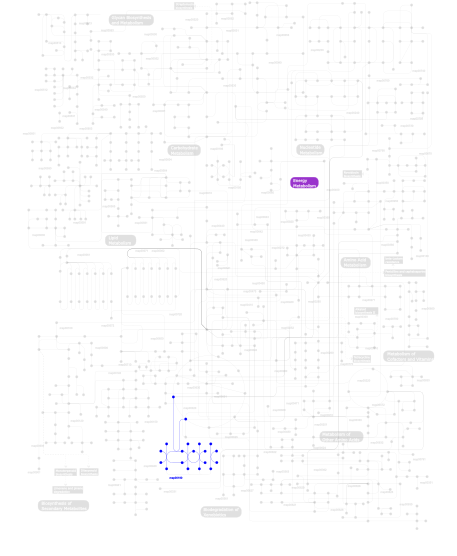
Click the image to view the interactive version of the map in iPath% proteins involved KEGG pathway ID Description 16.55 map05030 Amyotrophic lateral sclerosis (ALS) 16.55 map04210 Apoptosis 10.79 map05222 Small cell lung cancer 10.07 map05210 Colorectal cancer 6.47 map05212 Pancreatic cancer 6.47 map04630 Jak-STAT signaling pathway 6.47 map05220 Chronic myeloid leukemia 5.76 map05040 Huntington's disease 5.76 map04115 p53 signaling pathway 4.32 map05060 Prion disease 4.32 map04510 Focal adhesion 4.32 map05215 Prostate cancer 1.44 map01040 Biosynthesis of unsaturated fatty acids 0.72  map00190
Oxidative phosphorylation This information is based on mapping of SMART genomic protein database to KEGG orthologous groups. Percentage points are related to the number of proteins with BCL domain which could be assigned to a KEGG orthologous group, and not all proteins containing BCL domain. Please note that proteins can be included in multiple pathways, ie. the numbers above will not always add up to 100%.
- Structure (3D structures containing this domain)
3D Structures of BCL domains in PDB
PDB code Main view Title 1af3 
RAT BCL-XL AN APOPTOSIS INHIBITORY PROTEIN 1bxl 
STRUCTURE OF BCL-XL/BAK PEPTIDE COMPLEX, NMR, MINIMIZED AVERAGE STRUCTURE 1f16 
SOLUTION STRUCTURE OF A PRO-APOPTOTIC PROTEIN BAX 1g5j 
COMPLEX OF BCL-XL WITH PEPTIDE FROM BAD 1g5m 
HUMAN BCL-2, ISOFORM 1 1gjh 
HUMAN BCL-2, ISOFORM 2 1k3k 
Solution Structure of a Bcl-2 Homolog from Kaposi's Sarcoma Virus 1lxl 
NMR STRUCTURE OF BCL-XL, AN INHIBITOR OF PROGRAMMED CELL DEATH, MINIMIZED AVERAGE STRUCTURE 1maz 
X-RAY STRUCTURE OF BCL-XL, AN INHIBITOR OF PROGRAMMED CELL DEATH 1mk3 
SOLUTION STRUCTURE OF HUMAN BCL-W PROTEIN 1o0l 
THE STRUCTURE OF BCL-W REVEALS A ROLE FOR THE C-TERMINAL RESIDUES IN MODULATING BIOLOGICAL ACTIVITY 1ohu 
Structure of Caenorhabditis elegans CED-9 1pq0 
Crystal structure of mouse Bcl-xl 1pq1 
Crystal structure of Bcl-xl/Bim 1r2d 
Structure of Human Bcl-XL at 1.95 Angstroms 1r2e 
Human Bcl-XL containing a Glu to Leu mutation at position 92 1r2g 
Human Bcl-XL containing a Phe to Trp mutation at position 97 1r2h 
Human Bcl-XL containing an Ala to Leu mutation at position 142 1r2i 
Human Bcl-XL containing a Phe to Leu mutation at position 146 1ty4 
Crystal structure of a CED-9/EGL-1 complex 1wsx 
Solution structure of MCL-1 1ysg 
Solution Structure of the Anti-apoptotic Protein Bcl-xL in Complex with ""SAR by NMR"" Ligands 1ysi 
Solution structure of the anti-apoptotic protein Bcl-xL in complex with an acyl-sulfonamide-based ligand 1ysn 
Solution structure of the anti-apoptotic protein Bcl-xL complexed with an acyl-sulfonamide-based ligand 1ysw 
Solution structure of the anti-apoptotic protein Bcl-2 complexed with an acyl-sulfonamide-based ligand 1yxm 
Crystal structure of peroxisomal trans 2-enoyl CoA reductase 1zy3 
Structural model of complex of Bcl-w protein with Bid BH3-peptide 2a5y 
Structure of a CED-4/CED-9 complex 2b48 
Bcl-XL 3D Domain Swapped Dimer 2bzw 
The crystal structure of BCL-XL in complex with full-length BAD 2ims 
The X-ray Structure of a Bak Homodimer Reveals an Inhibitory Zinc Binding Site 2imt 
The X-ray Structure of a Bak Homodimer Reveals an Inhibitory Zinc Binding Site 2jcn 
The crystal structure of BAK1 - a mitochondrial apoptosis regulator 2jm6 
Solution structure of MCL-1 complexed with NOXAB 2k7w 
BAX Activation is Initiated at a Novel Interaction Site 2kbw 
Solution Structure of human Mcl-1 complexed with human Bid_BH3 peptide 2kua 
Solution structure of a divergent Bcl-2 protein 2lp8 
SOLUTION STRUCTURE OF AN APOPTOSIS ACTIVATING PHOTOSWITCHABLE BAK PEPTIDE BOUND to BCL-XL 2lpc 
NMR STRUCTURE of Bcl-XL 2lr1 
Structural Mechanism for Bax Inhibition by Cytomegalovirus Protein vMIA 2m03 
Solution structure of BCL-xL determined with selective isotope labelling of I,L,V sidechains 2m04 
Solution structure of BCL-xL in complex with PUMA BH3 peptide 2m5b 
The NMR structure of the BID-BAK complex 2me8 
Solution Structure of BCL-xL in its p53-bound conformation determined with selective isotope labelling of I,L,V sidechains 2me9 
Solution structure of BCL-xL containing the alpha1-alpha2 disordered loop determined with selective isotope labelling of I,L,V sidechains 2mej 
Solution Structure of the Complex Between BCL-xL and the p53 Core Domain determined with PRE restraints 2mhs 
NMR Structure of human Mcl-1 2nl9 
Crystal structure of the Mcl-1:Bim BH3 complex 2nla 
Crystal structure of the Mcl-1:mNoxaB BH3 complex 2o1y 
Solution structure of the anti-apoptotic protein Bcl-xL in complex with an acyl-sulfonamide-based ligand 2o21 
Solution structure of the anti-apoptotic protein Bcl-2 in complex with an acyl-sulfonamide-based ligand 2o22 
Solution structure of the anti-apoptotic protein Bcl-2 in complex with an acyl-sulfonamide-based ligand 2o2f 
Solution structure of the anti-apoptotic protein Bcl-2 in complex with an acyl-sulfonamide-based ligand 2o2m 
Solution structure of the anti-apoptotic protein Bcl-xL in complex with an acyl-sulfonamide-based ligand 2o2n 
Solution structure of the anti-apoptotic protein Bcl-xL in complex with an acyl-sulfonamide-based ligand 2p1l 
Structure of the Bcl-XL:Beclin 1 complex 2pon 
Solution structure of the Bcl-xL/Beclin-1 complex 2pqk 
X-ray crystal structure of human Mcl-1 in complex with Bim BH3 2roc 
Solution structure of Mcl-1 Complexed with Puma 2rod 
Solution Structure of MCL-1 Complexed with NoxaA 2vm6 
HUMAN BCL-2A1 in complex with BIM 2vof 
Structure of mouse A1 bound to the Puma BH3-domain 2vog 
Structure of mouse A1 bound to the Bmf BH3-domain 2voh 
Structure of mouse A1 bound to the Bak BH3-domain 2voi 
Structure of mouse A1 bound to the Bid BH3-domain 2w3l 
Crystal Structure of Chimaeric Bcl2-xL and Phenyl Tetrahydroisoquinoline Amide Complex 2xa0 
Crystal structure of BCL-2 in complex with a BAX BH3 peptide 2y6w 
Structure of a Bcl-w dimer 2yj1 
Puma BH3 foldamer in complex with Bcl-xL 2yq6 
Structure of Bcl-xL bound to BimSAHB 2yq7 
Structure of Bcl-xL bound to BimLOCK 2yv6 
Crystal structure of human Bcl-2 family protein Bak 2yxj 
Crystal structure of Bcl-xL in complex with ABT-737 3cva 
Human Bcl-xL containing a Trp to Ala mutation at position 137 3d7v 
Crystal structure of Mcl-1 in complex with an Mcl-1 selective BH3 ligand 3fdl 
Bim BH3 peptide in complex with Bcl-xL 3fdm 
alpha/beta foldamer in complex with Bcl-xL 3i1h 
Crystal structure of human BFL-1 in complex with BAK BH3 peptide 3ihc 
Crystal structure of mouse Bcl-xl (wt) at pH 5.0 3ihd 
Crystal structure of mouse Bcl-xl mutant (Y101A) at pH 5.0 3ihe 
Crystal structure of mouse Bcl-xl mutant (F105A) at pH 6.0 3ihf 
Crystal structure of mouse Bcl-xl mutant (R139A) at pH 5.0 3iig 
Crystal structure of mouse Bcl-xl mutant (F105A) at pH 5.0 3iih 
Crystal structure of mouse Bcl-xl (wt) at pH 6.0 3ilb 
Crystal structure of mouse Bcl-xl mutant (R139A) at pH 6.0 3ilc 
Crystal structure of mouse Bcl-xl mutant (Y101A) at pH 6.0 3inq 
Crystal structure of BCL-XL in complex with W1191542 3io8 
BimL12F in complex with Bcl-xL 3io9 
BimL12Y in complex with Mcl-1 3kj0 
Mcl-1 in complex with Bim BH3 mutant I2dY 3kj1 
Mcl-1 in complex with Bim BH3 mutant I2dA 3kj2 
Mcl-1 in complex with Bim BH3 mutant F4aE 3kz0 
MCL-1 complex with MCL-1-specific selected peptide 3mk8 
The MCL-1 BH3 Helix is an Exclusive MCL-1 Inhibitor and Apoptosis Sensitizer 3mqp 
Crystal Structure of human BFL-1 in complex with NOXA BH3 peptide, Northeast Structural Genomics Consortium Target HR2930 3pk1 
Crystal structure of Mcl-1 in complex with the BaxBH3 domain 3pl7 
Crystal structure of Bcl-xL in complex with the BaxBH3 domain 3qbr 
BakBH3 in complex with sjA 3qkd 
Crystal structure of Bcl-xL in complex with a Quinazoline sulfonamide inhibitor 3r85 
Crystal structure of human SOUL BH3 domain in complex with Bcl-xL 3sp7 
Crystal Structure of Bcl-xL bound to BM903 3spf 
Crystal Structure of Bcl-xL bound to BM501 3wix 
Crystal structure of Mcl-1 in complex with compound 4 3wiy 
Crystal structure of Mcl-1 in complex with compound 10 3wiz 
Crystal structure of Bcl-xL in complex with compound 10 3zk6 
Crystal structure of Bcl-xL in complex with inhibitor (Compound 2). 3zln 
Crystal structure of Bcl-xL in complex with inhibitor (Compound 3). 3zlo 
Crystal structure of Bcl-xL in complex with inhibitor (Compound 6). 3zlr 
Crystal structure of Bcl-xL in complex with inhibitor (WEHI-539). 4a1u 
Crystal structure of alpha-beta-foldamer 2c in complex with Bcl-xL 4a1w 
Crystal structure of alpha-beta foldamer 4c in complex with Bcl-xL 4aq3 
HUMAN BCL-2 WITH PHENYLACYLSULFONAMIDE INHIBITOR 4b4s 
Crystal Structure of a pro-survival Bcl-2:Bim BH3 complex 4bd2 
Bax domain swapped dimer in complex with BidBH3 4bd6 
Bax domain swapped dimer in complex with BaxBH3 4bd7 
Bax domain swapped dimer induced by octylmaltoside 4bd8 
Bax domain swapped dimer induced by BimBH3 with CHAPS 4bdu 
Bax BH3-in-Groove dimer (GFP) 4bpi 
Mcl-1 bound to alpha beta Puma BH3 peptide 2 4bpj 
Mcl-1 bound to alpha beta Puma BH3 peptide 3 4bpk 
Bcl-xL bound to alpha beta Puma BH3 peptide 5 4c52 
Crystal structure of Bcl-xL in complex with benzoylurea compound (39b) 4c5d 
Crystal structure of Bcl-xL in complex with benzoylurea compound (42) 4cim 
4CIM 4cin 
4CIN 4ehr 
Crystal structure of Bcl-Xl complex with 4-(5-butyl-3-(hydroxymethyl)-1-phenyl-1h-pyrazol-4-yl)-3-(3,4-dihydro-2(1h)-isoquinolinylcarbonyl)-n-((2-(trimethylsilyl)ethyl)sulfonyl)benzamide 4g35 
Mcl-1 in complex with a biphenyl cross-linked Noxa peptide. 4hnj 
Crystallographic structure of BCL-xL domain-swapped dimer in complex with PUMA BH3 peptide at 2.9A resolution 4hw2 
Discovery of potent Mcl-1 inhibitors using fragment-based methods and structure-based design 4hw3 
Discovery of potent Mcl-1 inhibitors using fragment-based methods and structure-based design 4hw4 
Discovery of potent Mcl-1 inhibitors using fragment-based methods and structure-based design 4ieh 
Crystal Structure of human Bcl-2 in complex with a small molecule inhibitor targeting Bcl-2 BH3 domain interactions 4k5a 
Co-crystallization with conformation-specific designed ankyrin repeat proteins explains the conformational flexibility of BCL-W 4k5b 
Co-crystallization with conformation-specific designed ankyrin repeat proteins explains the conformational flexibility of BCL-W 4lvt 
Bcl_2-Navitoclax (ABT-263) Complex 4lxd 
Bcl_2-Navitoclax Analog (without Thiophenyl) Complex 4man 
Bcl_2-Navitoclax Analog (with Indole) Complex 4oq5 
Crystal Structure of Human MCL-1 Bound to Inhibitor 4-(4-methylnaphthalen-1-yl)-2-{[(4-phenoxyphenyl)sulfonyl]amino}benzoic acid 4oq6 
Crystal Structure of Human MCL-1 Bound to Inhibitor 4-hydroxy-4'-propylbiphenyl-3-carboxylic acid 4ppi 
4PPI 4qnq 
4QNQ 4qve 
4QVE 4qvf 
4QVF 4qvx 
4QVX 4s0o 
4S0O 4s0p 
4S0P 4tuh 
4TUH 4u2u 
4U2U 4u2v 
4U2V 4wgi 
4WGI 4wmr 
4WMR 4wms 
4WMS 4wmt 
4WMT 4wmu 
4WMU 4wmv 
4WMV 4wmw 
4WMW 4wmx 
4WMX 4yj4 
4YJ4 4yk9 
4YK9 4z9v 
4Z9V 4zbf 
4ZBF 4zbi 
4ZBI 4zeq 
4ZEQ 4zie 
4ZIE 4zif 
4ZIF 4zig 
4ZIG 4zih 
4ZIH 4zii 
4ZII 5agw 
5AGW 5agx 
5AGX 5c3f 
5C3F 5c3g 
5C3G 5c6h 
5C6H 5fc4 
5FC4 5fcg 
5FCG 5fdo 
5FDO 5fdr 
5FDR 5fmi 
5FMI 5fmj 
5FMJ 5fmk 
5FMK 5jsb 
5JSB 5jsn 
5JSN 5ktg 
5KTG 5lof 
5LOF - Links (links to other resources describing this domain)
-
PROSITE BCL2_FAMILY PFAM Bcl-2