
GuKcGuanylate kinase homologues. |
|---|
| SMART accession number: | SM00072 |
|---|---|
| Description: | Active enzymes catalyze ATP-dependent phosphorylation of GMP to GDP. Structure resembles that of adenylate kinase. So-called membrane-associated guanylate kinase homologues (MAGUKs) do not possess guanylate kinase activities; instead at least some possess protein-binding functions. |
| Interpro abstract (IPR008145): | This entry represents a domain found in guanylate kinase ( EC 2.7.4.8 ) and in L-type calcium channel. Guanylate kinase ( EC 2.7.4.8 ) (GK) [ (PUBMED:1314905) ] catalyzes the ATP-dependent phosphorylation of GMP into GDP. It is essential for recycling GMP and indirectly, cGMP. In prokaryotes (such as Escherichia coli), lower eukaryotes (such as yeast) and in vertebrates, GK is a highly conserved monomeric protein of about 200 amino acids. GK has been shown [ (PUBMED:1310897) (PUBMED:8097461) (PUBMED:1329277) ] to be structurally similar to protein A57R (or SalG2R) from various strains of Vaccinia virus. L-type calcium channnels are formed from different alpha-1 subunit isoforms that determine the pharmacological properties of the channel, since they form the drug binding domain. Other properties, such as gating voltage-dependence, G protein modulation and kinase susceptibility, are influenced by alpha-2, delta and beta subunits. |
| Family alignment: |
There are 42084 GuKc domains in 42042 proteins in SMART's nrdb database.
Click on the following links for more information.
- Evolution (species in which this domain is found)
-
Taxonomic distribution of proteins containing GuKc domain.
This tree includes only several representative species. The complete taxonomic breakdown of all proteins with GuKc domain is also avaliable.
Click on the protein counts, or double click on taxonomic names to display all proteins containing GuKc domain in the selected taxonomic class.
- Literature (relevant references for this domain)
-
Primary literature is listed below; Automatically-derived, secondary literature is also avaliable.
- Kawabe H, Hata Y, Takeuchi M, Ide N, Mizoguchi A, Takai Y
- nArgBP2, a novel neural member of ponsin/ArgBP2/vinexin family that interacts with synapse-associated protein 90/postsynaptic density-95-associated protein (SAPAP).
- J Biol Chem. 1999; 274: 30914-8
- Display abstract
Postsynaptic density (PSD)-95/synapse-associated protein (SAP) 90 and synaptic scaffolding molecule (S-SCAM) are synaptic membrane-associated guanylate kinases. Both the proteins interact with SAP90/PSD-95-associated protein (SAPAP) (also called guanylate kinase-associated protein/Dlg-associated protein). SAPAP is a protein highly enriched in the PSD fraction and may link PSD-95/SAP90 and S-SCAM to Triton X-100-insoluble structures. We found here a novel SAPAP-interacting protein, which was specifically expressed in neural tissue and was present in the postsynaptic density fraction in brain. This protein had a sorbin homology domain in the N terminus, a zinc finger motif in the middle region, and three src homology (SH) 3 domains in the C terminus and was homologous to the ponsin/ArgBP2/vinexin family proteins. We named this protein nArgBP2 because it was the most homologous to ArgBP2. nArgBP2 is a neural member of a growing family of SH3-containing proteins. nArgBP2 bound to the proline-rich region of SAPAP via its third SH3 domain and was coimmunoprecipitated with SAPAP from the extract of rat brain. Furthermore, nArgBP2 was colocalized with SAPAP at synapses in cerebellum. nArgBP2 bound to not only SAPAP but also vinculin and l-afadin, known to bind to ponsin and vinexin. nArgBP2 may be implicated in the protein network around SAPAP in the PSD.
- Kuhlendahl S, Spangenberg O, Konrad M, Kim E, Garner CC
- Functional analysis of the guanylate kinase-like domain in the synapse-associated protein SAP97.
- Eur J Biochem. 1998; 252: 305-13
- Display abstract
SAP97 is a membrane cytoskeletal protein localized at the presynaptic nerve terminals of type 1 asymmetric synapses. It has been implicated in the assembly of synapses and in particular in the localization and clustering of ion channels. The C-terminal GK domain of SAP97 shares a high degree of sequence similarity with low-molecular-mass guanylate kinases. These enzymes are involved in the guanine nucleotide metabolic cycle and in the maintenance of GTP/GDP pools required for example in Ras-mediated cell signaling. It has therefore been hypothesized that SAP97 plays an essential role in cellular signaling by regulating the guanine nucleotide pools at synaptic junctions. Here, we test this hypothesis by assessing whether the GK domain in SAP97 encodes an authentic guanylate kinase. We show that the GK domain in and of itself does not encode an active guanylate kinase, that it cannot be activated by its binding partner GKAP and that flanking regions are not acting as inhibitory regulators for enzymatic activity. Thus, it would appear that the GK domain of SAP97 is not involved in the metabolism of guanine nucleotides required for signaling events.
- Rizo J, Sudhof TC
- C2-domains, structure and function of a universal Ca2+-binding domain.
- J Biol Chem. 1998; 273: 15879-82
- Kim E et al.
- GKAP, a novel synaptic protein that interacts with the guanylate kinase-like domain of the PSD-95/SAP90 family of channel clustering molecules.
- J Cell Biol. 1997; 136: 669-78
- Display abstract
The molecular mechanisms underlying the organization of ion channels and signaling molecules at the synaptic junction are largely unknown. Recently, members of the PSD-95/SAP90 family of synaptic MAGUK (membrane-associated guanylate kinase) proteins have been shown to interact, via their NH2-terminal PDZ domains, with certain ion channels (NMDA receptors and K+ channels), thereby promoting the clustering of these proteins. Although the function of the NH2-terminal PDZ domains is relatively well characterized, the function of the Src homology 3 (SH3) domain and the guanylate kinase-like (GK) domain in the COOH-terminal half of PSD-95 has remained obscure. We now report the isolation of a novel synaptic protein, termed GKAP for guanylate kinase-associated protein, that binds directly to the GK domain of the four known members of the mammalian PSD-95 family. GKAP shows a unique domain structure and appears to be a major constituent of the postsynaptic density. GKAP colocalizes and coimmunoprecipitates with PSD-95 in vivo, and coclusters with PSD-95 and K+ channels/NMDA receptors in heterologous cells. Given their apparent lack of guanylate kinase enzymatic activity, the fact that the GK domain can act as a site for protein-protein interaction has implications for the function of diverse GK-containing proteins (such as p55, ZO-1, and LIN-2/CASK).
- Ponting CP, Phillips C, Davies KE, Blake DJ
- PDZ domains: targeting signalling molecules to sub-membranous sites.
- Bioessays. 1997; 19: 469-79
- Display abstract
PDZ (also called DHR or GLGF) domains are found in diverse membrane-associated proteins including members of the MAGUK family of guanylate kinase homologues, several protein phosphatases and kinases, neuronal nitric oxide synthase, and several dystrophin-associated proteins, collectively known as syntrophins. Many PDZ domain-containing proteins appear to be localised to highly specialised submembranous sites, suggesting their participation in cellular junction formation, receptor or channel clustering, and intracellular signalling events. PDZ domains of several MAGUKs interact with the C-terminal polypeptides of a subset of NMDA receptor subunits and/or with Shaker-type K+ channels. Other PDZ domains have been shown to bind similar ligands of other transmembrane receptors. Recently, the crystal structures of PDZ domains, with and without ligand, have been determined. These demonstrate the mode of ligand-binding and the structural bases for sequence conservation among diverse PDZ domains.
- Kim SK
- Tight junctions, membrane-associated guanylate kinases and cell signaling.
- Curr Opin Cell Biol. 1995; 7: 641-9
- Display abstract
Proteins that define a new family, termed membrane-associated guanylate kinases, have recently been identified as structural components of epithelial tight junctions and neuronal synapses, and as cell signaling proteins in Drosophila and Caenorhabditis elegans. In particular, the lin-2 gene has been shown to encode a membrane-associated guanylate kinase and to act in the let-23 receptor tyrosine kinase/let-60 ras signaling pathway that controls vulval induction in C. elegans. The combined data from recent biochemical and genetic analyses of membrane-associated guanylate kinases suggest that certain tight junction proteins can play an important role in cell signaling pathways. One possibility is that asymmetric segregation of signaling receptors to the basolateral membrane domain of polarized epithelial cells is crucial for proper cell signaling, and that membrane-associated guanylate kinases may be required for this localization.
- Stehle T, Schulz GE
- Three-dimensional structure of the complex of guanylate kinase from yeast with its substrate GMP.
- J Mol Biol. 1990; 211: 249-54
- Display abstract
The enzyme guanylate kinase was isolated from baker's yeast and crystallized as a complex with its substrate GMP. The crystal structure was solved by multiple isomorphous replacement, solvent-flattening, restrained least-squares refinement, and simulated annealing. The current R-factor is 28.9% at a resolution of 2.0 A. The model is given as a backbone tracing, the GMP binding site is shown in atomic detail. In its major domain (residues 1 to 32 and 82 to 186), the chain fold is closely similar to the adenylate kinases, while the minor domain (residues 33 to 81) differs grossly from the 3-helix fold of the adenylate kinases. Structural homology and mechanistical similarity allow us to assign the AMP site of the adenylate kinases on the basis of the GMP site.
- Metabolism (metabolic pathways involving proteins which contain this domain)
-
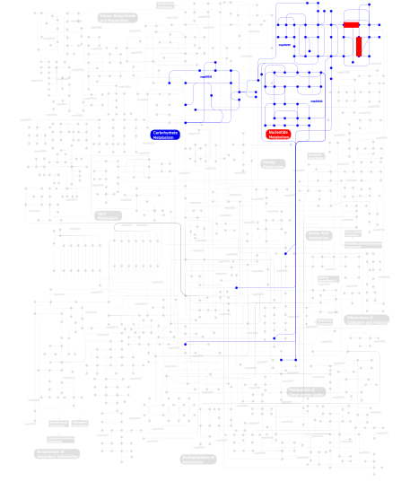
Click the image to view the interactive version of the map in iPath% proteins involved KEGG pathway ID Description 74.96  map00230
Purine metabolism 8.73 map04530 Tight junction 6.59 map00030Pentose phosphate pathway 4.61 map04010 MAPK signaling pathway 1.32 map05050 Dentatorubropallidoluysian atrophy (DRPLA) 1.15 map04520 Adherens junction 1.15 map05120 Epithelial cell signaling in Helicobacter pylori infection 1.15 map04540 Gap junction 0.16 map05219 Bladder cancer 0.16 map00240Pyrimidine metabolism This information is based on mapping of SMART genomic protein database to KEGG orthologous groups. Percentage points are related to the number of proteins with GuKc domain which could be assigned to a KEGG orthologous group, and not all proteins containing GuKc domain. Please note that proteins can be included in multiple pathways, ie. the numbers above will not always add up to 100%.
- Structure (3D structures containing this domain)
3D Structures of GuKc domains in PDB
PDB code Main view Title 1ex6 
CRYSTAL STRUCTURE OF UNLIGANDED FORM OF GUANYLATE KINASE FROM YEAST 1ex7 
CRYSTAL STRUCTURE OF YEAST GUANYLATE KINASE IN COMPLEX WITH GUANOSINE-5'-MONOPHOSPHATE 1gky 
REFINED STRUCTURE OF THE COMPLEX BETWEEN GUANYLATE KINASE AND ITS SUBSTRATE GMP AT 2.0 ANGSTROMS RESOLUTION 1jxm 
CRYSTAL STRUCTURE OF THE GMP BOUND SH3-HOOK-GK FRAGMENT OF PSD-95 1jxo 
Crystal Structure of the SH3-HOOK-GK Fragment of PSD-95 1kgd 
Crystal Structure of the Guanylate Kinase-like Domain of Human CASK 1kjw 
SH3-Guanylate Kinase Module from PSD-95 1lvg 
Crystal structure of mouse guanylate kinase in complex with GMP and ADP 1s4q 
Crystal Structure of Guanylate Kinase from Mycobacterium tuberculosis (Rv1389) 1s96 
The 2.0 A X-ray structure of Guanylate Kinase from E.coli 1t0h 
Crystal structure of the Rattus norvegicus voltage gated calcium channel beta subunit isoform 2a 1t0j 
Crystal structure of a complex between voltage-gated calcium channel beta2a subunit and a peptide of the alpha1c subunit 1t3l 
Structural Analysis of the Voltage-Dependent Calcium Channel Beta Subunit Functional Core in Complex with Alpha1 Interaction Domain 1t3s 
Structural Analysis of the Voltage-Dependent Calcium Channel Beta Subunit Functional Core 1vyt 
beta3 subunit complexed with aid 1vyu 
Beta3 subunit of Voltage-gated Ca2+-channel 1vyv 
beta4 subunit of Ca2+ channel 1z6g 
Crystal structure of guanylate kinase from Plasmodium falciparum 1z8f 
Guanylate Kinase Double Mutant A58C, T157C from Mycobacterium tuberculosis (Rv1389) 1znw 
Crystal Structure Of Unliganded Form Of Mycobacterium tuberculosis Guanylate Kinase 1znx 
Crystal Structure Of Mycobacterium tuberculosis Guanylate Kinase In Complex With GMP 1zny 
Crystal Structure Of Mycobacterium tuberculosis Guanylate Kinase In Complex With GDP 1znz 
Crystal Structure Of The Reduced Form Of Mycobacterium tuberculosis Guanylate Kinase In Complex With GDP 2an9 
Crystal Structure Of Oligomeric E.coli Guanylate Kinase In Complex With GDP 2anb 
Crystal Structure Of Oligomeric E.coli Guanylate Kinase In Complex With GMP 2anc 
Crystal Structure Of Unliganded Form Of Oligomeric E.coli Guanylate Kinase 2f3r 
Crystal Structure Of E.coli Guanylate Kinase In Complex With Ap5G 2f3t 
Crystal Structure Of E.coli Guanylate Kinase In Complex With Ganciclovir monophosphate 2j41 
Crystal structure of Staphylococcus aureus guanylate monophosphate kinase 2qor 
Crystal structure of Plasmodium vivax guanylate kinase 2xkx 
Single particle analysis of PSD-95 in negative stain 3jbr 
3JBR 3kfv 
Crystal structure of the SH3-kinase fragment of tight junction protein 3 (TJP3) in apo-form 3lh5 
Crystal Structure of the SH3-Guanylate kinase core domain of ZO-1 3lnc 
Crystal structure of guanylate kinase from Anaplasma phagocytophilum 3ney 
Crystal structure of the kinase domain of MPP1/p55 3shw 
Crystal structure of ZO-1 PDZ3-SH3-Guk supramodule complex with Connexin-45 peptide 3tau 
Crystal Structure of a Putative Guanylate Monophosphaste Kinase from Listeria monocytogenes EGD-e 3tr0 
Structure of guanylate kinase (gmk) from Coxiella burnetii 3tsw 
crystal structure of the PDZ3-SH3-GUK core module of Human ZO-1 3tsz 
crystal structure of PDZ3-SH3-GUK core module from human ZO-1 in complex with 12mer peptide from human JAM-A cytoplasmic tail 3tvt 
Structural basis for Discs Large interaction with Pins 3uat 
Guanylate Kinase Domains of the MAGUK Family Scaffold Proteins as Specific Phospho-Protein Binding Modules 3w9y 
Crystal structure of the human DLG1 guanylate kinase domain 3wp0 
Crystal structure of Dlg GK in complex with a phosphor-Lgl2 peptide 3wp1 
Phosphorylation-dependent interaction between tumor suppressors Dlg and Lgl 4dex 
Crystal structure of the Voltage Dependent Calcium Channel beta-2 Subunit in Complex With The CaV2.2 I-II Linker. 4dey 
Crystal structure of the Voltage Dependent Calcium Channel beta-2 Subunit in Complex With The CaV1.2 I-II Linker. 4f4j 
Conversion of the enzyme guanylate kinase into a mitotic spindle orienting protein by a single mutation that inhibits gmp- induced closing 4qrh 
4QRH 4wsi 
4WSI 4zw2 
4ZW2 5b64 
5B64 5gjv 
5GJV 5gjw 
5GJW - Links (links to other resources describing this domain)
-
PROSITE GuKc_DOMAIN INTERPRO IPR008145