LRRCTLeucine rich repeat C-terminal domain |
|---|
| SMART accession number: | SM00082 |
|---|---|
| Description: | - |
| Interpro abstract (IPR000483): | Leucine-rich repeats (LRR, see IPR001611 ) consist of 2-45 motifs of 20-30 amino acids in length that generally folds into an arc or horseshoe shape [ (PUBMED:14747988) ]. LRRs occur in proteins ranging from viruses to eukaryotes, and appear to provide a structural framework for the formation of protein-protein interactions [ (PUBMED:11751054) ]. Proteins containing LRRs include tyrosine kinase receptors, cell-adhesion molecules, virulence factors, and extracellular matrix-binding glycoproteins, and are involved in a variety of biological processes, including signal transduction, cell adhesion, DNA repair, recombination, transcription, RNA processing, disease resistance, apoptosis, and the immune response. LRRs are often flanked by cysteine-rich domains: an N-terminal LRR domain ( IPR000372 ) and a C-terminal LRR domain. This entry represents the C-terminal LRR domain. |
| Family alignment: |
There are 43815 LRRCT domains in 36442 proteins in SMART's nrdb database.
Click on the following links for more information.
- Evolution (species in which this domain is found)
-
Taxonomic distribution of proteins containing LRRCT domain.
This tree includes only several representative species. The complete taxonomic breakdown of all proteins with LRRCT domain is also avaliable.
Click on the protein counts, or double click on taxonomic names to display all proteins containing LRRCT domain in the selected taxonomic class.
- Literature (relevant references for this domain)
-
Primary literature is listed below; Automatically-derived, secondary literature is also avaliable.
- Kobe B, Deisenhofer J
- A structural basis of the interactions between leucine-rich repeats and protein ligands.
- Nature. 1995; 374: 183-6
- Display abstract
The leucine-rich repeat is a recently characterized structural motif used in molecular recognition processes as diverse as signal transduction, cell adhesion, cell development, DNA repair and RNA processing. We present here the crystal structure at 2.5 A resolution of the complex between ribonuclease A and ribonuclease inhibitor, a protein built entirely of leucine-rich repeats. The unusual non-globular structure of ribonuclease inhibitor, its solvent-exposed parallel beta-sheet and the conformational flexibility of the structure are used in the interaction; they appear to be the principal reasons for the effectiveness of leucine-rich repeats as protein-binding motifs. The structure can serve as a model for the interactions of other proteins containing leucine-rich repeats with their ligands.
- Kobe B, Deisenhofer J
- The leucine-rich repeat: a versatile binding motif.
- Trends Biochem Sci. 1994; 19: 415-21
- Display abstract
Leucine-rich repeats are short sequence motifs present in a number of proteins with diverse functions and cellular locations. All proteins containing these repeats are thought to be involved in protein-protein interactions. The crystal structure of ribonuclease inhibitor protein has revealed that leucine-rich repeats correspond to beta-alpha structural units. These units are arranged so that they form a parallel beta-sheet with one surface exposed to solvent, so that the protein acquires an unusual, nonglobular shape. These two features may be responsible for the protein-binding functions of proteins containing leucine-rich repeats.
- Disease (disease genes where sequence variants are found in this domain)
-
SwissProt sequences and OMIM curated human diseases associated with missense mutations within the LRRCT domain.
Protein Disease Platelet glycoprotein Ib alpha chain (P07359) (SMART) OMIM:231200: Bernard-Soulier syndrome - Metabolism (metabolic pathways involving proteins which contain this domain)
-

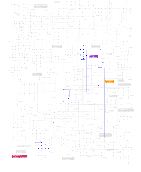
Click the image to view the interactive version of the map in iPath% proteins involved KEGG pathway ID Description 36.42 map04620 Toll-like receptor signaling pathway 16.18 map04512 ECM-receptor interaction 13.29 map04360 Axon guidance 12.14 map04640 Hematopoietic cell lineage 6.94 map04010 MAPK signaling pathway 4.05 map04510 Focal adhesion 2.89 map05216 Thyroid cancer 2.89 map04210 Apoptosis 1.73  map00680
Methane metabolism 1.73  map00940
Phenylpropanoid biosynthesis 1.73  map00360
Phenylalanine metabolism This information is based on mapping of SMART genomic protein database to KEGG orthologous groups. Percentage points are related to the number of proteins with LRRCT domain which could be assigned to a KEGG orthologous group, and not all proteins containing LRRCT domain. Please note that proteins can be included in multiple pathways, ie. the numbers above will not always add up to 100%.
- Structure (3D structures containing this domain)
3D Structures of LRRCT domains in PDB
PDB code Main view Title 1gwb 
structure of glycoprotein 1b 1m0z 
Crystal Structure of the von Willebrand Factor Binding Domain of Glycoprotein Ib alpha 1m10 
Crystal structure of the complex of Glycoprotein Ib alpha and the von Willebrand Factor A1 Domain 1ook 
Crystal Structure of the Complex of Platelet Receptor GPIb-alpha and Human alpha-Thrombin 1ozn 
1.5A Crystal Structure of the Nogo Receptor Ligand Binding Domain Reveals a Convergent Recognition Scaffold Mediating Inhibition of Myelination 1p8t 
Crystal structure of Nogo-66 Receptor 1p8v 
CRYSTAL STRUCTURE OF THE COMPLEX OF PLATELET RECEPTOR GPIB-ALPHA AND ALPHA-THROMBIN AT 2.6A 1p9a 
Crystal Structure of N-Terminal Domain of Human Platelet Receptor Glycoprotein Ib-alpha at 1.7 Angstrom Resolution 1qyy 
Crystal Structure of N-Terminal Domain of Human Platelet Receptor Glycoprotein Ib-alpha at 2.8 Angstrom Resolution 1sq0 
Crystal Structure of the Complex of the Wild-type Von Willebrand Factor A1 domain and Glycoprotein Ib alpha at 2.6 Angstrom Resolution 1u0n 
The ternary von Willebrand Factor A1-glycoprotein Ibalpha-botrocetin complex 1w8a 
Third LRR domain of Drosophila Slit 1ziw 
Human Toll-like Receptor 3 extracellular domain structure 2a0z 
The molecular structure of toll-like receptor 3 ligand binding domain 2id5 
Crystal Structure of the Lingo-1 Ectodomain 2ifg 
Structure of the extracellular segment of human TRKA in complex with nerve growth factor 2o6q 
Structural diversity of the hagfish Variable Lymphocyte Receptors A29 2o6r 
Structural diversity of the hagfish Variable Lymphocyte Receptors B61 2o6s 
Structural diversity of the hagfish Variable Lymphocyte Receptors B59 2r9u 
Crystal Structure of Lamprey Variable Lymphocyte Receptor 2913 Ectodomain 2v70 
Third LRR domain of human Slit2 2v9s 
Second LRR domain of human Slit2 2v9t 
Complex between the second LRR domain of Slit2 and The first Ig domain from Robo1 2wfh 
The Human Slit 2 Dimerization Domain D4 2xot 
Crystal structure of neuronal leucine rich repeat protein AMIGO-1 2z62 
Crystal structure of the TV3 hybrid of human TLR4 and hagfish VLRB.61 2z63 
Crystal structure of the TV8 hybrid of human TLR4 and hagfish VLRB.61 2z64 
Crystal structure of mouse TLR4 and mouse MD-2 complex 2z65 
Crystal structure of the human TLR4 TV3 hybrid-MD-2-Eritoran complex 2z66 
Crystal structure of the VT3 hybrid of human TLR4 and hagfish VLRB.61 2z7x 
Crystal structure of the TLR1-TLR2 heterodimer induced by binding of a tri-acylated lipopeptide 2z80 
Crystal structure of the TLR1-TLR2 heterodimer induced by binding of a tri-acylated lipopeptide 2z81 
Crystal structure of the TLR1-TLR2 heterodimer induced by binding of a tri-acylated lipopeptide 2z82 
Crystal structure of the TLR1-TLR2 heterodimer induced by binding of a tri-acylated lipopeptide 3a79 
Crystal structure of TLR2-TLR6-Pam2CSK4 complex 3a7b 
Crystal structure of TLR2-Streptococcus Pneumoniae lipoteichoic acid complex 3a7c 
Crystal structure of TLR2-PE-DTPA complex 3b2d 
Crystal structure of human RP105/MD-1 complex 3cig 
Crystal structure of mouse TLR3 ectodomain 3ciy 
Mouse Toll-like receptor 3 ectodomain complexed with double-stranded RNA 3e6j 
Crystal Structure of Variable Lymphocyte Receptor (VLR) RBC36 in Complex with H-trisaccharide 3fxi 
Crystal structure of the human TLR4-human MD-2-E.coli LPS Ra complex 3g39 
Structure of a lamprey variable lymphocyte receptor 3g3a 
Structure of a lamprey variable lymphocyte receptor in complex with a protein antigen 3j0a 
Homology model of human Toll-like receptor 5 fitted into an electron microscopy single particle reconstruction 3kj4 
Structure of rat Nogo receptor bound to 1D9 antagonist antibody 3p72 
structure of platelet Glycoprotein 1b alpha with a bound peptide inhibitor 3pmh 
Mechanism of Sulfotyrosine-Mediated Glycoprotein Ib Interaction with Two Distinct alpha-Thrombin Sites 3rez 
glycoprotein GPIb variant 3rfe 
Crystal structure of glycoprotein GPIb ectodomain 3rfj 
Design of a binding scaffold based on variable lymphocyte receptors of jawless vertebrates by module engineering 3rfs 
Design of a binding scaffold based on variable lymphocyte receptors of jawless vertebrates by module engineering 3rg1 
Crystal structure of the RP105/MD-1 complex 3t6q 
Crystal structure of mouse RP105/MD-1 complex 3ul7 
Crystal structure of the TV3 mutant F63W 3ul8 
Crystal structure of the TV3 mutant V134L 3ul9 
structure of the TV3 mutant M41E 3ula 
Crystal structure of the TV3 mutant F63W-MD-2-Eritoran complex 3ulu 
Structure of quaternary complex of human TLR3ecd with three Fabs (Form1) 3ulv 
Structure of quaternary complex of human TLR3ecd with three Fabs (Form2) 3v44 
Crystal structure of the N-terminal fragment of zebrafish TLR5 3v47 
Crystal structure of the N-tetminal fragment of zebrafish TLR5 in complex with Salmonella flagellin 3vq1 
Crystal structure of mouse TLR4/MD-2/lipid IVa complex 3vq2 
Crystal structure of mouse TLR4/MD-2/LPS complex 3w3g 
Crystal structure of human TLR8 (unliganded form) 3w3j 
Crystal structure of human TLR8 in complex with CL097 3w3k 
Crystal structure of human TLR8 in complex with CL075 3w3l 
Crystal structure of human TLR8 in complex with Resiquimod (R848) crystal form 1 3w3m 
Crystal structure of human TLR8 in complex with Resiquimod (R848) crystal form 2 3w3n 
Crystal structure of human TLR8 in complex with Resiquimod (R848) crystal form 3 3wn4 
Crystal structure of human TLR8 in complex with DS-877 3zyi 
NetrinG2 in complex with NGL2 3zyj 
NetrinG1 in complex with NGL1 3zyn 
Crystal structure of the N-terminal leucine rich repeats of Netrin-G Ligand-3 3zyo 
Crystal structure of the N-terminal leucine rich repeats and immunoglobulin domain of netrin-G ligand-3 4arn 
Crystal structure of the N-terminal domain of Drosophila Toll receptor 4arr 
Crystal structure of the N-terminal domain of Drosophila Toll receptor with the magic triangle I3C 4bv4 
Structure and allostery in Toll-Spatzle recognition 4c2a 
Crystal Structure of High-Affinity von Willebrand Factor A1 domain with R1306Q and I1309V Mutations in Complex with High Affinity GPIb alpha 4c2b 
Crystal Structure of High-Affinity von Willebrand Factor A1 domain with Disulfide Mutation in Complex with High Affinity GPIb alpha 4cnc 
Crystal structure of human 5T4 (Wnt-activated inhibitory factor 1, Trophoblast glycoprotein) 4cnm 
Crystal structure of human 5T4 (Wnt-activated inhibitory factor 1, Trophoblast glycoprotein) 4g8a 
Crystal structure of human TLR4 polymorphic variant D299G and T399I in complex with MD-2 and LPS 4j4l 
Modular evolution and design of the protein binding interface 4lxr 
Structure of the Toll - Spatzle complex, a molecular hub in Drosophila development and innate immunity 4lxs 
Structure of the Toll - Spatzle complex, a molecular hub in Drosophila development and innate immunity (glycosylated form) 4oqt 
LINGO-1/Li81 Fab complex 4p8s 
Crystal structure of Nogo-receptor-2 4p91 
Crystal structure of the nogo-receptor-2 (27-330) 4pbv 
4PBV 4pbw 
4PBW 4q3g 
4Q3G 4q3i 
4Q3I 4qbz 
4QBZ 4qc0 
4QC0 4qdh 
4QDH 4qxe 
4QXE 4qxf 
4QXF 4r07 
4R07 4r08 
4R08 4r09 
4R09 4r0a 
4R0A 4r6a 
4R6A 4rca 
4RCA 4rcw 
4RCW 4u7l 
4U7L 4uip 
4UIP 4v2c 
4V2C 4v2d 
4V2D 4v2e 
4V2E 4xsq 
4XSQ 4y61 
4Y61 4yeb 
4YEB 5awa 
5AWA 5awb 
5AWB 5awc 
5AWC 5awd 
5AWD 5az5 
5AZ5 5cmn 
5CMN 5cmp 
5CMP 5d3i 
5D3I 5ftt 
5FTT 5ftu 
5FTU 5gmf 
5GMF 5gmg 
5GMG 5gmh 
5GMH 5gs0 
5GS0 5gs2 
5GS2 5hdh 
5HDH 5ijb 
5IJB 5ijc 
5IJC 5ijd 
5IJD - Links (links to other resources describing this domain)
-
INTERPRO IPR000483

