
TRASHmetallochaperone-like domain |
|---|
| SMART accession number: | SM00746 |
|---|---|
| Description: | - |
| Interpro abstract (IPR011017): | TRASH domain contains a well-conserved cysteine motif that may be involved in metal coordination. TRASH is encoded by multiple prokaryotic genomes and is present in transcriptional regulators, cation-transporting ATPases and hydrogenases, and is also present as a stand-alone module. The observed domain associations and conserved genome context of TRASH-encoding genes in prokaryotic genomes suggest that TRASH constitutes a novel component in metal trafficking and heavy-metal resistance. The precise role of the multiple copies of TRASH that are present in vertebrate proteins remains to be elucidated [ (PUBMED:12713899) ]. |
| Family alignment: |
There are 24136 TRASH domains in 7020 proteins in SMART's nrdb database.
Click on the following links for more information.
- Evolution (species in which this domain is found)
-
Taxonomic distribution of proteins containing TRASH domain.
This tree includes only several representative species. The complete taxonomic breakdown of all proteins with TRASH domain is also avaliable.
Click on the protein counts, or double click on taxonomic names to display all proteins containing TRASH domain in the selected taxonomic class.
- Literature (relevant references for this domain)
-
Primary literature is listed below; Automatically-derived, secondary literature is also avaliable.
- Ettema TJ, Huynen MA, de Vos WM, van der Oost J
- TRASH: a novel metal-binding domain predicted to be involved in heavy-metal sensing, trafficking and resistance.
- Trends Biochem Sci. 2003; 28: 170-3
- Display abstract
We describe a previously undetected domain - TRASH - containing a well-conserved cysteine motif that we anticipate to be involved in metal coordination. TRASH is encoded by multiple prokaryotic genomes and is present in transcriptional regulators, cation-transporting ATPases and hydrogenases, and is also present as a stand-alone module. The observed domain associations and conserved genome context of TRASH-encoding genes in prokaryotic genomes suggest that TRASH constitutes a novel component in metal trafficking and heavy-metal resistance. The role of the multiple copies of TRASH that are present in vertebrate proteins remains to be elucidated.
- Metabolism (metabolic pathways involving proteins which contain this domain)
-
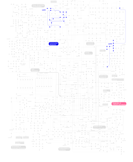
Click the image to view the interactive version of the map in iPath% proteins involved KEGG pathway ID Description 85.71 map03010 Ribosome 7.14  map00500
Starch and sucrose metabolism 7.14 map00790Folate biosynthesis This information is based on mapping of SMART genomic protein database to KEGG orthologous groups. Percentage points are related to the number of proteins with TRASH domain which could be assigned to a KEGG orthologous group, and not all proteins containing TRASH domain. Please note that proteins can be included in multiple pathways, ie. the numbers above will not always add up to 100%.
- Structure (3D structures containing this domain)
3D Structures of TRASH domains in PDB
PDB code Main view Title 1ffk 
CRYSTAL STRUCTURE OF THE LARGE RIBOSOMAL SUBUNIT FROM HALOARCULA MARISMORTUI AT 2.4 ANGSTROM RESOLUTION 1jj2 
Fully Refined Crystal Structure of the Haloarcula marismortui Large Ribosomal Subunit at 2.4 Angstrom Resolution 1k73 
Co-crystal Structure of Anisomycin Bound to the 50S Ribosomal Subunit 1k8a 
Co-crystal structure of Carbomycin A bound to the 50S ribosomal subunit of Haloarcula marismortui 1k9m 
Co-crystal structure of tylosin bound to the 50S ribosomal subunit of Haloarcula marismortui 1kc8 
Co-crystal Structure of Blasticidin S Bound to the 50S Ribosomal Subunit 1kd1 
Co-crystal Structure of Spiramycin bound to the 50S Ribosomal Subunit of Haloarcula marismortui 1kqs 
The Haloarcula marismortui 50S Complexed with a Pretranslocational Intermediate in Protein Synthesis 1m1k 
Co-crystal structure of azithromycin bound to the 50S ribosomal subunit of Haloarcula marismortui 1m90 
Co-crystal structure of CCA-Phe-caproic acid-biotin and sparsomycin bound to the 50S ribosomal subunit 1ml5 
Structure of the E. coli ribosomal termination complex with release factor 2 1n8r 
Structure of large ribosomal subunit in complex with virginiamycin M 1nji 
Structure of chloramphenicol bound to the 50S ribosomal subunit 1q7y 
Crystal Structure of CCdAP-Puromycin bound at the Peptidyl transferase center of the 50S ribosomal subunit 1q81 
Crystal Structure of minihelix with 3' puromycin bound to A-site of the 50S ribosomal subunit. 1q82 
Crystal Structure of CC-Puromycin bound to the A-site of the 50S ribosomal subunit 1q86 
Crystal structure of CCA-Phe-cap-biotin bound simultaneously at half occupancy to both the A-site and P-site of the the 50S ribosomal Subunit. 1qvf 
Structure of a deacylated tRNA minihelix bound to the E site of the large ribosomal subunit of Haloarcula marismortui 1qvg 
Structure of CCA oligonucleotide bound to the tRNA binding sites of the large ribosomal subunit of Haloarcula marismortui 1s72 
REFINED CRYSTAL STRUCTURE OF THE HALOARCULA MARISMORTUI LARGE RIBOSOMAL SUBUNIT AT 2.4 ANGSTROM RESOLUTION 1vq4 
The structure of the transition state analogue ""DAA"" bound to the large ribosomal subunit of Haloarcula marismortui 1vq5 
The structure of the transition state analogue ""RAA"" bound to the large ribosomal subunit of haloarcula marismortui 1vq6 
The structure of c-hpmn and CCA-PHE-CAP-BIO bound to the large ribosomal subunit of haloarcula marismortui 1vq7 
The structure of the transition state analogue ""DCA"" bound to the large ribosomal subunit of haloarcula marismortui 1vq8 
The structure of CCDA-PHE-CAP-BIO and the antibiotic sparsomycin bound to the large ribosomal subunit of haloarcula marismortui 1vq9 
The structure of CCA-PHE-CAP-BIO and the antibiotic sparsomycin bound to the large ribosomal subunit of haloarcula marismortui 1vqk 
The structure of CCDA-PHE-CAP-BIO bound to the a site of the ribosomal subunit of haloarcula marismortui 1vql 
The structure of the transition state analogue ""DCSN"" bound to the large ribosomal subunit of haloarcula marismortui 1vqm 
The structure of the transition state analogue ""DAN"" bound to the large ribosomal subunit of haloarcula marismortui 1vqn 
The structure of CC-HPMN AND CCA-PHE-CAP-BIO bound to the large ribosomal subunit of haloarcula marismortui 1vqo 
The structure of CCPMN bound to the large ribosomal subunit haloarcula marismortui 1vqp 
The structure of the transition state analogue ""RAP"" bound to the large ribosomal subunit of haloarcula marismortui 1w2b 
Trigger Factor ribosome binding domain in complex with 50S 1yhq 
Crystal Structure Of Azithromycin Bound To The G2099A Mutant 50S Ribosomal Subunit Of Haloarcula Marismortui 1yi2 
Crystal Structure Of Erythromycin Bound To The G2099A Mutant 50S Ribosomal Subunit Of Haloarcula Marismortui 1yij 
Crystal Structure Of Telithromycin Bound To The G2099A Mutant 50S Ribosomal Subunit Of Haloarcula Marismortui 1yit 
Crystal Structure Of Virginiamycin M and S Bound To The 50S Ribosomal Subunit Of Haloarcula Marismortui 1yj9 
Crystal Structure Of The Mutant 50S Ribosomal Subunit Of Haloarcula Marismortui Containing a three residue deletion in L22 1yjn 
Crystal Structure Of Clindamycin Bound To The G2099A Mutant 50S Ribosomal Subunit Of Haloarcula Marismortui 1yjw 
Crystal Structure Of Quinupristin Bound To The G2099A Mutant 50S Ribosomal Subunit Of Haloarcula Marismortui 2das 
Solution structure of TRASH domain of zinc finger MYM-type protein 5 2otj 
13-deoxytedanolide bound to the large subunit of Haloarcula marismortui 2otl 
Girodazole bound to the large subunit of Haloarcula marismortui 2qa4 
A more complete structure of the the L7/L12 stalk of the Haloarcula marismortui 50S large ribosomal subunit 2qex 
Negamycin Binds to the Wall of the Nascent Chain Exit Tunnel of the 50S Ribosomal Subunit 3cc2 
The Refined Crystal Structure of the Haloarcula Marismortui Large Ribosomal Subunit at 2.4 Angstrom Resolution with rrnA Sequence for the 23S rRNA and Genome-derived Sequences for r-Proteins 3cc4 
Co-crystal Structure of Anisomycin Bound to the 50S Ribosomal Subunit 3cc7 
Structure of Anisomycin resistant 50S Ribosomal Subunit: 23S rRNA mutation C2487U 3cce 
Structure of Anisomycin resistant 50S Ribosomal Subunit: 23S rRNA mutation U2535A 3ccj 
Structure of Anisomycin resistant 50S Ribosomal Subunit: 23S rRNA mutation C2534U 3ccl 
Structure of Anisomycin resistant 50S Ribosomal Subunit: 23S rRNA mutation U2535C. Density for Anisomycin is visible but not included in model. 3ccm 
Structure of Anisomycin resistant 50S Ribosomal Subunit: 23S rRNA mutation G2611U 3ccq 
Structure of Anisomycin resistant 50S Ribosomal Subunit: 23S rRNA mutation A2488U 3ccr 
Structure of Anisomycin resistant 50S Ribosomal Subunit: 23S rRNA mutation A2488C. Density for anisomycin is visible but not included in the model. 3ccs 
Structure of Anisomycin resistant 50S Ribosomal Subunit: 23S rRNA mutation G2482A 3ccu 
Structure of Anisomycin resistant 50S Ribosomal Subunit: 23S rRNA mutation G2482C 3ccv 
Structure of Anisomycin resistant 50S Ribosomal Subunit: 23S rRNA mutation G2616A 3cd6 
Co-cystal of large Ribosomal Subunit mutant G2616A with CC-Puromycin 3cma 
The structure of CCA and CCA-Phe-Cap-Bio bound to the large ribosomal subunit of Haloarcula marismortui 3cme 
The Structure of CA and CCA-PHE-CAP-BIO Bound to the Large Ribosomal Subunit of Haloarcula Marismortui 3cpw 
The structure of the antibiotic LINEZOLID bound to the large ribosomal subunit of HALOARCULA MARISMORTUI 3cxc 
The structure of an enhanced oxazolidinone inhibitor bound to the 50S ribosomal subunit of H. marismortui 3g4s 
Co-crystal structure of Tiamulin bound to the large ribosomal subunit 3g6e 
Co-crystal structure of Homoharringtonine bound to the large ribosomal subunit 3g71 
Co-crystal structure of Bruceantin bound to the large ribosomal subunit 3i55 
Co-crystal structure of Mycalamide A Bound to the Large Ribosomal Subunit 3i56 
Co-crystal structure of Triacetyloleandomcyin Bound to the Large Ribosomal Subunit 3j7o 
3J7O 3j7p 
3J7P 3j7q 
3J7Q 3j7r 
3J7R 3j92 
3J92 3jag 
3JAG 3jah 
3JAH 3jai 
3JAI 3jaj 
3JAJ 3jan 
3JAN 3jct 
3JCT 3ow2 
Crystal Structure of Enhanced Macrolide Bound to 50S Ribosomal Subunit 4adx 
The Cryo-EM Structure of the Archaeal 50S Ribosomal Subunit in Complex with Initiation Factor 6 4d5y 
4D5Y 4d67 
4D67 4ug0 
4UG0 4ujc 
4UJC 4ujd 
4UJD 4uje 
4UJE 4v3p 
4V3P 4v42 
4V42 4v4n 
4V4N 4v4p 
4V4P 4v4r 
4V4R 4v4s 
4V4S 4v4t 
4V4T 4v5z 
4V5Z 4v6u 
4V6U 4v6w 
4V6W 4v6x 
4V6X 4v7e 
4V7E 4v7f 
4V7F 4v9f 
4V9F 5aj0 
5AJ0 5fl8 
5FL8 5jcs 
5JCS 5lzs 
5LZS 5lzt 
5LZT 5lzu 
5LZU 5lzv 
5LZV 5lzw 
5LZW 5lzx 
5LZX 5lzy 
5LZY 5lzz 
5LZZ - Links (links to other resources describing this domain)
-
PFAM YHS domain INTERPRO IPR011017