
WWDomain with 2 conserved Trp (W) residues |
|---|
| SMART accession number: | SM00456 |
|---|---|
| Description: | Also known as the WWP or rsp5 domain. Binds proline-rich polypeptides. |
| Interpro abstract (IPR001202): | Synonym(s): Rsp5 or WWP domain The WW domain is a short conserved region in a number of unrelated proteins, which folds as a stable, triple stranded beta-sheet. This short domain of approximately 40 amino acids, may be repeated up to four times in some proteins [ (PUBMED:7846762) (PUBMED:7802651) (PUBMED:7828727) (PUBMED:7641887) ]. The name WW or WWP derives from the presence of two signature tryptophan residues that are spaced 20-23 amino acids apart and are present in most WW domains known to date, as well as that of a conserved Pro. The WW domain binds to proteins with particular proline-motifs, [AP]-P-P-[AP]-Y, and/or phosphoserine- phosphothreonine-containing motifs [ (PUBMED:7644498) (PUBMED:11911877) ]. It is frequently associated with other domains typical for proteins in signal transduction processes. A large variety of proteins containing the WW domain are known. These include; dystrophin, a multidomain cytoskeletal protein; utrophin, a dystrophin-like protein of unknown function; vertebrate YAP protein, substrate of an unknown serine kinase; Mus musculus (Mouse) NEDD-4, involved in the embryonic development and differentiation of the central nervous system; Saccharomyces cerevisiae (Baker's yeast) RSP5, similar to NEDD-4 in its molecular organisation; Rattus norvegicus (Rat) FE65, a transcription-factor activator expressed preferentially in liver; Nicotiana tabacum (Common tobacco) DB10 protein, amongst others. |
| GO function: | protein binding (GO:0005515) |
| Family alignment: |
There are 67566 WW domains in 38399 proteins in SMART's nrdb database.
Click on the following links for more information.
- Evolution (species in which this domain is found)
-
Taxonomic distribution of proteins containing WW domain.
This tree includes only several representative species. The complete taxonomic breakdown of all proteins with WW domain is also avaliable.
Click on the protein counts, or double click on taxonomic names to display all proteins containing WW domain in the selected taxonomic class.
- Cellular role (predicted cellular role)
-
Cellular role: interaction, signalling
Binding / catalysis: protein-binding, polyproline-binding - Literature (relevant references for this domain)
-
Primary literature is listed below; Automatically-derived, secondary literature is also avaliable.
- Bedford MT, Reed R, Leder P
- WW domain-mediated interactions reveal a spliceosome-associated protein that binds a third class of proline-rich motif: the proline glycine and methionine-rich motif.
- Proc Natl Acad Sci U S A. 1998; 95: 10602-7
- Display abstract
Pre-mRNA splicing requires the bridging of the 5' and 3' ends of the intron. In yeast, this bridging involves interactions between the WW domains in the splicing factor PRP40 and a proline-rich domain in the branchpoint binding protein, BBP. Using a proline-rich domain derived from formin (a product of the murine limb deformity locus), we have identified a family of murine formin binding proteins (FBP's), each of which contains one or more of a special class of tyrosine-rich WW domains. Two of these WW domains, in the proteins FBP11 and FBP21, are strikingly similar to those found in the yeast splicing factor PRP40. We show that FBP21 is present in highly purified spliceosomal complex A, is associated with U2 snRNPs, and colocalizes with splicing factors in nuclear speckle domains. Moreover, FBP21 interacts directly with the U1 snRNP protein U1C, the core snRNP proteins SmB and SmB', and the branchpoint binding protein SF1/mBBP. Thus, FBP21 may play a role in cross-intron bridging of U1 and U2 snRNPs in the mammalian A complex.
- Neele DM, Kaptein A, Huisman H, deWit EC, Princen HM
- No effect of fibrates on synthesis of apolipoprotein(a) in primary cultures of cynomolgus monkey and human hepatocytes: apolipoprotein A-I synthesis increased.
- Biochem Biophys Res Commun. 1998; 244: 374-8
- Display abstract
Fibrates have been shown to decrease plasma levels of triglyceride-rich lipoproteins and LDL and to increase HDL. Data on the effect of fibrates on lipoprotein(a) levels in man are not consistent. Because lp(a) levels in vivo are mainly regulated at synthesis level, we studied the effect of fibrates on the synthesis of apolipoprotein(a) (apo(a)) in primary cultures of cynomolgus monkey and human hepatocytes. Furthermore, we assessed the effect of fibrates on apolipoprotein A-I (apo A-I) synthesis and investigated whether different fibrates have different effects on the apo(a) and apo A-I synthesis. The addition of gemfibrozil to cultures of monkey and human hepatocytes had no effect on apo(a) synthesis, but resulted in a dose- and time-dependent increase of apo A-I synthesis and mRNA. In simian hepatocytes maximal stimulation was 2.5-fold after incubation for 72 h with 1.0 mM gemfibrozil, whereas apo A-I synthesis was induced 1.8- and 2.0-fold by using 0.1 mM and 0.3 mM, respectively. Similar results were obtained by using human hepatocytes; apo(a) synthesis remained unchanged, while apo A-I secretion was 2.0-fold increased at 1 mM gemfibrozil. Other fibrates like bezafibrate, clofibrate and clofibric acid did not change apo(a) synthesis either. In contrast, they enhanced the synthesis of apo A-I (1.5-, 1.8- and 1.8-fold, respectively), although less potently than gemfibrozil. We conclude that fibrates have no effect on apolipoprotein(a) synthesis in monkey and human hepatocytes and that these drugs induce apo A-I synthesis.
- Rotin D
- WW (WWP) domains: from structure to function.
- Curr Top Microbiol Immunol. 1998; 228: 115-33
- DiFiore PP, Pelicci PG, Sorkin A
- EH: a novel protein-protein interaction domain potentially involved in intracellular sorting.
- Trends Biochem Sci. 1997; 22: 411-3
- Ermekova KS et al.
- The WW domain of neural protein FE65 interacts with proline-rich motifs in Mena, the mammalian homolog of Drosophila enabled.
- J Biol Chem. 1997; 272: 32869-77
- Display abstract
The neural protein FE65 contains two types of protein-protein interaction modules: one WW binding domain and two phosphotyrosine binding domains. The carboxyl-terminal phosphotyrosine binding domain of FE65 interacts in vivo with the beta-amyloid precursor protein, which is implicated in Alzheimer disease. To understand the function of this adapter protein, we identified binding partners for the FE65 WW domain. Proline-rich sequences sharing a proline-proline-leucine-proline core motif were recovered by screening expression libraries for ligands of the FE65 WW domain. Five proteins of molecular masses 60, 75, 80, 140, and 200 kDa could be purified from mouse brain lysates by affinity to the FE65 WW domain. We identified two of these five proteins as the 80- and 140-kDa isoforms encoded by Mena, the mammalian homolog of the Drosophila Enabled gene. Using the SPOTs technique of peptide synthesis, we identified the sequences in Mena that interact with the FE65 WW domain and found that they contain the signature proline-proline-leucine-proline motif. Finally, we demonstrated that Mena binds to FE65 in vivo by coimmunoprecipitation assay from COS cell extracts. The specificity of the Mena-FE65 WW domain association was confirmed by competition assays. Further characterization of the FE65-Mena complex may identify a physiological role for these proteins in beta-amyloid precursor protein biogenesis and may help in understanding the mechanism of molecular changes that underlie Alzheimer disease.
- Gavva NR, Gavva R, Ermekova K, Sudol M, Shen CJ
- Interaction of WW domains with hematopoietic transcription factor p45/NF-E2 and RNA polymerase II.
- J Biol Chem. 1997; 272: 24105-8
- Display abstract
NF-E2 is an erythroid-specific transcription factor required for expression of several erythroid-specific genes. By Far-Western blotting and yeast two-hybrid assay, we demonstrate that p45, the large subunit of NF-E2, is capable of binding to a specific set of WW domain-containing proteins, including the ubiquitin ligase hRPF1. This binding is mediated through the interaction between the WW domains and a PY motif located within the amino-terminal region of p45. Interestingly, the carboxyl-terminal domain of mammalian RNA polymerase II binds a similar set of WW domains to which p45 interacts with. We discuss the data in terms of possible new pathways through which the processes of transcriptional regulation by NF-E2 could be regulated in erythroid and megakaryote cells.
- Macias MJ et al.
- Structure of the WW domain of a kinase-associated protein complexed with a proline-rich peptide.
- Nature. 1996; 382: 646-9
- Display abstract
The WW domain is a new protein module with two highly conserved tryptophans that binds proline-rich peptide motifs in vitro. It is present in a number of signalling and regulatory proteins, often in several copies. Here we investigate the solution structure of the WW domain of human YAP65 (for Yes kinase-associated protein) in complex with proline-rich peptides containing the core motif PPxY. The structure of the domain with the bound peptide GTPPPPYTVG is a slightly curved, three-stranded, antiparallel beta-sheet. Two prolines pack against the first tryptophan, forming a hydrophobic buckle on the convex side of the sheet. The concave side has three exposed hydrophobic residues (tyrosine, tryptophan and leucine) which form the binding site for the ligand. A non-conserved isoleucine in the amino-terminal flanking region covers a hydrophobic patch and stabilizes the WW domain of human YAP65 in vitro. The structure of the WW domain differs from that of the SH3 domain and reveals a new design for a protein module that uses stacked aromatic surface residues to arrange a binding site for proline-rich peptides.
- Ponting CP, Blake DJ, Davies KE, Kendrick-Jones J, Winder SJ
- ZZ and TAZ: new putative zinc fingers in dystrophin and other proteins.
- Trends Biochem Sci. 1996; 21: 11-13
- Hofmann K, Bucher P
- The rsp5-domain is shared by proteins of diverse functions.
- FEBS Lett. 1995; 358: 153-7
- Display abstract
A novel, unusually small, and highly conserved domain of modular intracellular proteins is described. The domain was first recognized as three repeats in the yeast rsp5 gene product and named thereafter. The rsp5 protein is thought to interact with nuclear proteins but also contains a C2 domain typical for cytoplasmic proteins. Further analyses revealed several additional occurrences of this domain in diverse protein classes, including cytoplasmic signal transduction proteins, gene products interacting with the transcription machinery, structural proteins like dystrophin, and a putative RNA helicase.
- Sudol M et al.
- Characterization of the mammalian YAP (Yes-associated protein) gene and its role in defining a novel protein module, the WW domain.
- J Biol Chem. 1995; 270: 14733-41
- Display abstract
We report cDNA cloning and characterization of the human and mouse orthologs of the chicken YAP (Yes-associated protein) gene which encodes a novel protein that binds to the SH3 (Src homology 3) domain of the Yes proto-oncogene product. Sequence comparison between mouse, human, and chicken YAP proteins showed an inserted sequence in the mouse YAP that represented an imperfect repeat of an upstream sequence. Further analysis of this sequence revealed a putative protein module that is found in various structural, regulatory, and signaling molecules in yeast, nematode, and mammals including human dystrophin. Because one of the prominent features of this sequence motif is two tryptophans (W), we named it the WW domain (Bork, P., and Sudol, M. (1994) Trends Biochem. Sci. 19, 531-533). Since its delineation, more proteins have been shown to contain this domain, and we report here on the widespread distribution of the WW module and present a discussion of its possible function. We have also shown that the human YAP gene is well conserved among higher eukaryotes, but it may not be conserved in yeast. Its expression at the RNA level in adult human tissues is nearly ubiquitous, being relatively high in placenta, prostate, ovary, and testis, but is not detectable in peripheral blood leukocytes. Using fluorescence in situ hybridization on human metaphase chromosomes and by analyzing rodent-human hybrids by Southern blot hybridization and polymerase chain reaction amplification, we mapped the human YAP gene to chromosome band 11q13, a region to which the multiple endocrine neoplasia type 1 gene has been mapped.
- Andre B, Springael JY
- WWP, a new amino acid motif present in single or multiple copies in various proteins including dystrophin and the SH3-binding Yes-associated protein YAP65.
- Biochem Biophys Res Commun. 1994; 205: 1201-5
- Display abstract
A new repeating amino acid motif, which we called WWP, was found in several proteins of yeast, nematod or vertebrate origin. Among these are dystrophin, the product of the Duchenne muscular dystrophy locus, a protein (YAP65) which associates in vitro with the Src homology domain 3 (SH3) of the Yes proto-oncogene product, and a human putative GTPase-activating protein. As is the case for proteins which contain the SH2, SH3 and PH domains, at least some of the WWP-containing proteins appear to be signaling or cytoskeletal proteins, associated with plasma or organellar membranes, and specific protein-protein contacts are likely to be crucial to their function.
- Bork P, Sudol M
- The WW domain: a signalling site in dystrophin?
- Trends Biochem Sci. 1994; 19: 531-3
- Kobe B, Deisenhofer J
- The leucine-rich repeat: a versatile binding motif.
- Trends Biochem Sci. 1994; 19: 415-21
- Display abstract
Leucine-rich repeats are short sequence motifs present in a number of proteins with diverse functions and cellular locations. All proteins containing these repeats are thought to be involved in protein-protein interactions. The crystal structure of ribonuclease inhibitor protein has revealed that leucine-rich repeats correspond to beta-alpha structural units. These units are arranged so that they form a parallel beta-sheet with one surface exposed to solvent, so that the protein acquires an unusual, nonglobular shape. These two features may be responsible for the protein-binding functions of proteins containing leucine-rich repeats.
- Maclennan AJ, Shaw G
- A yeast SH2 domain.
- Trends Biochem Sci. 1993; 18: 464-5
- Metabolism (metabolic pathways involving proteins which contain this domain)
-
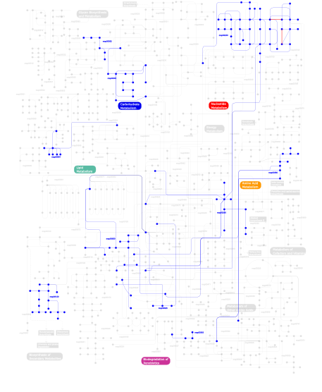
Click the image to view the interactive version of the map in iPath% proteins involved KEGG pathway ID Description 23.53 map04120 Ubiquitin mediated proteolysis 21.57 map05050 Dentatorubropallidoluysian atrophy (DRPLA) 15.69 map04530 Tight junction 10.78 map04350 TGF-beta signaling pathway 5.88 map05010 Alzheimer's disease 3.92 map04810 Regulation of actin cytoskeleton 1.96  map00260
Glycine, serine and threonine metabolism 1.96 map00625Tetrachloroethene degradation 1.96 map00650Butanoate metabolism 1.96 map00591Linoleic acid metabolism 1.96 map00052Galactose metabolism 1.96 map00120Bile acid biosynthesis 1.96 map00380Tryptophan metabolism 1.96 map00363Bisphenol A degradation 1.96 map00051Fructose and mannose metabolism 0.98 map00230Purine metabolism This information is based on mapping of SMART genomic protein database to KEGG orthologous groups. Percentage points are related to the number of proteins with WW domain which could be assigned to a KEGG orthologous group, and not all proteins containing WW domain. Please note that proteins can be included in multiple pathways, ie. the numbers above will not always add up to 100%.
- Structure (3D structures containing this domain)
3D Structures of WW domains in PDB
PDB code Main view Title 1e0l 
FBP28WW domain from Mus musculus 1e0m 
PROTOTYPE WW domain 1eg3 
STRUCTURE OF A DYSTROPHIN WW DOMAIN FRAGMENT IN COMPLEX WITH A BETA-DYSTROGLYCAN PEPTIDE 1eg4 
STRUCTURE OF A DYSTROPHIN WW DOMAIN FRAGMENT IN COMPLEX WITH A BETA-DYSTROGLYCAN PEPTIDE 1f8a 
STRUCTURAL BASIS FOR THE PHOSPHOSERINE-PROLINE RECOGNITION BY GROUP IV WW DOMAINS 1i5h 
SOLUTION STRUCTURE OF THE RNEDD4 WWIII DOMAIN-RENAC BP2 PEPTIDE COMPLEX 1i6c 
SOLUTION STRUCTURE OF PIN1 WW DOMAIN 1i8g 
SOLUTION STRUCTURE OF PIN1 WW DOMAIN COMPLEXED WITH CDC25 PHOSPHOTHREONINE PEPTIDE 1i8h 
SOLUTION STRUCTURE OF PIN1 WW DOMAIN COMPLEXED WITH HUMAN TAU PHOSPHOTHREONINE PEPTIDE 1jmq 
YAP65 (L30K mutant) WW domain in Complex with GTPPPPYTVG peptide 1k5r 
YAP65 WW domain S24-Amino-Ethylsulfanyl-Acetic Acid mutant 1k9q 
YAP65 WW domain complexed to N-(n-octyl)-GPPPY-NH2 1k9r 
YAP65 WW domain complexed to Acetyl-PLPPY 1nmv 
Solution structure of human Pin1 1o6w 
Solution Structure of the Prp40 WW Domain Pair of the Yeast Splicing Factor Prp40 1pin 
PIN1 PEPTIDYL-PROLYL CIS-TRANS ISOMERASE FROM HOMO SAPIENS 1tk7 
NMR structure of WW domains (WW3-4) from Suppressor of Deltex 1wmv 
Solution structure of the second WW domain of WWOX 1wr3 
Solution structure of the first WW domain of Nedd4-2 1wr4 
Solution structure of the second WW domain of Nedd4-2 1wr7 
Solution structure of the third WW domain of Nedd4-2 1yiu 
Itch E3 ubiquitin ligase WW3 domain 1ymz 
CC45, An Artificial WW Domain Designed Using Statistical Coupling Analysis 1yw5 
Peptidyl-prolyl isomerase ESS1 from Candida albicans 1ywi 
Structure of the FBP11WW1 domain complexed to the peptide APPTPPPLPP 1ywj 
Structure of the FBP11WW1 domain 1zcn 
human Pin1 Ng mutant 1zr7 
Solution structure of the first WW domain of FBP11 2djy 
Solution structure of Smurf2 WW3 domain-Smad7 PY peptide complex 2dk1 
Solution structure of WW domain in WW domain binding protein 4 (WBP-4) 2dk7 
Solution structure of WW domain in transcription elongation regulator 1 2dmv 
Solution structure of the second ww domain of Itchy homolog E3 ubiquitin protein ligase (Itch) 2dwv 
Solution structure of the second WW domain from mouse salvador homolog 1 protein (mWW45) 2dyf 
Solution structure of the first WW domain of FBP11 / HYPA (FBP11 WW1) complexed with a PL (PPLP) motif peptide ligand 2e45 
Solution structure of Fe65 WW domain 2ez5 
Solution Structure of the dNedd4 WW3* Domain- Comm LPSY Peptide Complex 2f21 
human Pin1 Fip mutant 2ho2 
Structure of human FE65-WW domain in complex with hMena peptide. 2idh 
Crystal Structure of human FE65 WW domain 2itk 
human Pin1 bound to D-PEPTIDE 2jmf 
Solution structure of the Su(dx) WW4- Notch PY peptide complex 2jo9 
Mouse Itch 3rd WW domain complex with the Epstein-Barr virus latent membrane protein 2A derived peptide EEPPPPYED 2joc 
Mouse Itch 3rd domain phosphorylated in T30 2jup 
FBP28WW2 domain in complex with the PPLIPPPP peptide 2jv4 
Structure Characterisation of PINA WW Domain and Comparison with other Group IV WW Domains, PIN1 and ESS1 2jx8 
Solution structure of hPCIF1 WW domain 2jxw 
Solution Structure of the Tandem WW Domains of FBP21 2kbu 
NMR solution structure of Pin1 WW domain mutant with beta turn mimic at position 12 2kcf 
The NMR solution structure of the isolated Apo Pin1 WW domain 2kpz 
Human NEDD4 3RD WW Domain Complex with The Human T-cell Leukemia virus 1 GAG-Pro poliprotein Derived Peptide SDPQIPPPYVEP 2kq0 
Human NEDD4 3rd WW Domain Complex with Ebola Zaire Virus Matrix Protein VP40 Derived Peptide ILPTAPPEYMEA 2kxq 
Solution Structure of Smurf2 WW2 and WW3 bound to Smad7 PY motif containing peptide 2kyk 
The sandwich region between two LMP2A PY motif regulates the interaction between AIP4WW2domain and PY motif 2l4j 
Yap ww2 2l5f 
Solution structure of the tandem WW domains from HYPA/FBP11 2laj 
Third WW domain of human Nedd4L in complex with doubly phosphorylated human smad3 derived peptide 2law 
Structure of the second WW domain from human YAP in complex with a human Smad1 derived peptide 2lax 
Structure of first WW domain of human YAP in complex with a human Smad1 doubly-phosphorilated derived peptide. 2lay 
Structure of the first WW domain of human YAP in complex with a phosphorylated human Smad1 derived peptide 2laz 
Structure of the first WW domain of human Smurf1 in complex with a mono-phosphorylated human Smad1 derived peptide 2lb0 
Structure of the first WW domain of human Smurf1 in complex with a di-phosphorylated human Smad1 derived peptide 2lb1 
Structure of the second domain of human Smurf1 in complex with a human Smad1 derived peptide 2lb2 
Structure of the second domain of human Nedd4L in complex with a phosphorylated pTPY motif derived from human Smad3 2lb3 
Structure of the WW domain of PIN1 in complex with a human phosphorylated Smad3 derived peptide 2ltv 
YAP WW2 in complex with a Smad7 derived peptide 2ltw 
YAP WW1 in complex with a Smad7 derived peptide 2ltx 
Smurf1 WW2 domain in complex with a Smad7 derived peptide 2lty 
NEDD4L WW2 domain in complex with a Smad7 derived peptide 2ltz 
Smurf2 WW3 domain in complex with a Smad7 derived peptide 2m3o 
Structure and dynamics of a human Nedd4 WW domain-ENaC complex 2m8i 
Structure of Pin1 WW domain 2m8j 
Structure of Pin1 WW domain phospho-mimic S16E 2m9e 
NMR solution structure of Pin1 WW domain mutant 5-1 2m9f 
NMR solution structure of Pin1 WW domain mutant 5-1g 2m9i 
NMR solution structure of Pin1 WW domain variant 6-1 2m9j 
NMR solution structure of Pin1 WW domain mutant 6-1g 2mdc 
2MDC 2mdi 
2MDI 2mdj 
2MDJ 2mpt 
2MPT 2mw9 
2MW9 2mwa 
2MWA 2mwb 
2MWB 2mwd 
2MWD 2mwe 
2MWE 2mwf 
2MWF 2n1o 
2N1O 2n4r 
2N4R 2n4s 
2N4S 2n4t 
2N4T 2n4u 
2N4U 2n4v 
2N4V 2n4w 
2N4W 2n8s 
2N8S 2n8t 
2N8T 2n8u 
2N8U 2nc3 
2NC3 2nc4 
2NC4 2nc5 
2NC5 2nc6 
2NC6 2nnt 
General structural motifs of amyloid protofilaments 2oei 
Crystal structure of human FE65-WW domain in complex with human Mena peptide 2op7 
WW4 2q5a 
human Pin1 bound to L-PEPTIDE 2rly 
FBP28WW2 domain in complex with PTPPPLPP peptide 2rm0 
FBP28WW2 domain in complex with a PPPLIPPPP peptide 2xp3 
DISCOVERY OF CELL-ACTIVE PHENYL-IMIDAZOLE PIN1 INHIBITORS BY STRUCTURE-GUIDED FRAGMENT EVOLUTION 2xp4 
DISCOVERY OF CELL-ACTIVE PHENYL-IMIDAZOLE PIN1 INHIBITORS BY STRUCTURE-GUIDED FRAGMENT EVOLUTION 2xp5 
DISCOVERY OF CELL-ACTIVE PHENYL-IMIDAZOLE PIN1 INHIBITORS BY STRUCTURE-GUIDED FRAGMENT EVOLUTION 2xp6 
DISCOVERY OF CELL-ACTIVE PHENYL-IMIDAZOLE PIN1 INHIBITORS BY STRUCTURE-GUIDED FRAGMENT EVOLUTION 2xp7 
DISCOVERY OF CELL-ACTIVE PHENYL-IMIDAZOLE PIN1 INHIBITORS BY STRUCTURE-GUIDED FRAGMENT EVOLUTION 2xp8 
DISCOVERY OF CELL-ACTIVE PHENYL-IMIDAZOLE PIN1 INHIBITORS BY STRUCTURE-GUIDED FRAGMENT EVOLUTION 2xp9 
DISCOVERY OF CELL-ACTIVE PHENYL-IMIDAZOLE PIN1 INHIBITORS BY STRUCTURE-GUIDED FRAGMENT EVOLUTION 2xpa 
DISCOVERY OF CELL-ACTIVE PHENYL-IMIDAZOLE PIN1 INHIBITORS BY STRUCTURE-GUIDED FRAGMENT EVOLUTION 2xpb 
DISCOVERY OF CELL-ACTIVE PHENYL-IMIDAZOLE PIN1 INHIBITORS BY STRUCTURE-GUIDED FRAGMENT EVOLUTION 2ysb 
Solution structure of the first WW domain from the mouse salvador homolog 1 protein (SAV1) 2ysc 
Solution structure of the WW domain from the human amyloid beta A4 precursor protein-binding family B member 3, APBB3 2ysd 
Solution structure of the first WW domain from the human membrane-associated guanylate kinase, WW and PDZ domain-containing protein 1. MAGI-1 2yse 
Solution structure of the second WW domain from the human membrane-associated guanylate kinase, WW and PDZ domain-containing protein 1. MAGI-1 2ysf 
Solution structure of the fourth WW domain from the human E3 ubiquitin-protein ligase Itchy homolog, ITCH 2ysg 
Solution structure of the WW domain from the human syntaxin-binding protein 4 2ysh 
Solution structure of the WW domain from the human growth-arrest-specific protein 7, GAS-7 2ysi 
Solution structure of the first WW domain from the mouse transcription elongation regulator 1, transcription factor CA150 2zaj 
Solution structure of the short-isoform of the second WW domain from the human membrane-associated guanylate kinase, WW and PDZ domain-containing protein 1 (MAGI-1) 2zqs 
Crystal structure of a mutant PIN1 PEPTIDYL-PROLYL CIS-TRANS ISOMERASE 2zqt 
Crystal structure of a mutant PIN1 PEPTIDYL-PROLYL CIS-TRANS ISOMERASE 2zqu 
Crystal structure of a mutant PIN1 PEPTIDYL-PROLYL CIS-TRANS ISOMERASE 2zqv 
Crystal structure of a mutant PIN1 PEPTIDYL-PROLYL CIS-TRANS ISOMERASE 2zr4 
Crystal structure of a mutant PIN1 peptidyl-prolyl cis-trans isomerase 2zr5 
Crystal structure of a mutant PIN1 peptidyl-prolyl cis-trans isomerase 2zr6 
Crystal structure of a mutant PIN1 peptidyl-prolyl cis-trans isomerase 3kab 
Structure-guided design of alpha-amino acid-derived Pin1 inhibitors 3kad 
Structure-guided design of alpha-amino acid-derived Pin1 inhibitors 3kaf 
Structure-guided design of alpha-amino acid-derived Pin1 inhibitors 3kag 
Structure-guided design of alpha-amino acid-derived Pin1 inhibitors 3kah 
Structure-guided design of alpha-amino acid-derived Pin1 inhibitors 3kai 
Structure-guided design of alpha-amino acid-derived Pin1 inhibitors 3kce 
Structure-guided design of alpha-amino acid-derived Pin1 inhibitors 3l4h 
Helical box domain and second WW domain of the human E3 ubiquitin-protein ligase HECW1 3le4 
Crystal structure of the DGCR8 dimerization domain 3ntp 
Human Pin1 complexed with reduced amide inhibitor 3odk 
Discovery of cell-active phenyl-imidazole Pin1 inhibitors by structure-guided fragment evolution 3olm 
Structure and Function of a Ubiquitin Binding Site within the Catalytic Domain of a HECT Ubiquitin Ligase 3oob 
Structural and functional insights of directly targeting Pin1 by Epigallocatechin-3-gallate 3tc5 
Selective targeting of disease-relevant protein binding domains by O-phosphorylated natural product derivatives 3tcz 
Human Pin1 bound to cis peptidomimetic inhibitor 3tdb 
Human Pin1 bound to trans peptidomimetic inhibitor 3wh0 
3WH0 4e5r 
Crystal Structure of Frog DGCR8 Dimerization Domain 4gwt 
Structure of racemic Pin1 WW domain cocrystallized with DL-malic acid 4gwv 
Structure of racemic Pin1 WW domain cocrystallized with tri-ammonium citrate 4lcd 
Structure of an Rsp5xUbxSna3 complex: Mechanism of ubiquitin ligation and lysine prioritization by a HECT E3 4n7f 
Crystal structure of 3rd WW domain of human Nedd4-1 4n7h 
Crystal Structure of the Complex of 3rd WW domain of Human Nedd4 and 1st PPXY Motif of ARRDC3 4qib 
4QIB 4rex 
4REX 4rof 
4ROF 4u84 
4U84 4u85 
4U85 4u86 
4U86 5aht 
5AHT 5b3z 
5B3Z 5cq2 
5CQ2 5dws 
5DWS 5dzd 
5DZD - Links (links to other resources describing this domain)
-
INTERPRO IPR001202 PFAM WW_rsp5_WWP PROSITE WW_DOMAIN_2