This domain conveys a zinc dependent phospholipase C activity (EC 3.1.4.3). It is found in a monomeric phospholipase C of Bacillus cereus as well as in the alpha toxin of Clostridium perfringens and Clostridium bifermentans, which is involved in haemolysis and cell rupture. It is also found in a lecithinase of Listeria monocytogenes, which is involved in breaking the 2-membrane vacuoles that surround the bacterium. Structure information: PDB 1ca1.
Bacillus cereus contains a monomeric phospholipase C EC 3.1.4.3 (PLC) of 245 amino-acid residues that binds three zinc ions [ (PUBMED:2493587) ]. Although PLC prefers to act on phosphatidylcholine, it also shows weak catalytic activity with sphingomyelin and phosphatidylinositol [ (PUBMED:2841128) ]. Sequence studies have shown the PLC protein to be similar to the following:
Alpha toxin from Clostridium perfringens and Clostridium bifermentans, which are zinc-dependent phospholipases C involved in haemolysis and cell rupture [ (PUBMED:2536355) ].
Lecithinase C from Listeria monocytogenes, which aids cell-to-cell spread by breaking down the 2-membrane vacuoles that surround the bacterium during transfer [ (PUBMED:1309513) ].
Each of these proteins is a zinc-dependent enzyme, binding 3 zinc ions per molecule [ (PUBMED:2111259) ]. The enzymes catalyse the conversion of phosphatidylcholine and water to 1,2-diacylglycerol and choline phosphate [ (PUBMED:2841128) (PUBMED:2536355) (PUBMED:2111259) ]. In B. cereus, there are nine residues known to be involved in binding the zinc ions: 5 His, 2 Asp, 1 Glu and 1 Trp. These residues are all conserved in the Clostridium alpha-toxin [ (PUBMED:9699639) ].
Site-directed mutagenesis of histidine residues in Clostridium perfringens alpha-toxin.
J Bacteriol. 1995; 177: 1179-85
Display abstract
Mutagenesis of H-68 or -148 in Clostridium perfringens alpha-toxin resulted in complete loss of hemolytic, phospholipase C, sphingomyelinase, and lethal activities of the toxin. These activities of the variant toxin at H-126 or -136 decreased by approximately 100-fold of the activities of the wild-type toxin. Mutation at H-46, -207, -212, or -241 showed no effect on the biological activities, indicating that these residues are not essential for these activities. The variant toxin at H-11 was not detected in culture supernatant and in cells of the transformant carrying the variant toxin gene. Wild-type toxin and the variant toxin at H-148 bound to erythrocytes in the presence of Ca2+; however, the variant toxins at H-68, -126, and -136 did not. Co2+ and Mn2+ ions stimulated binding of the variant toxin at H-68, -126, and -136 to membranes in the presence of Ca2+ and caused an increase in hemolytic activity. Wild-type toxin and the variant toxins at H-68, -126, and -136 contained two zinc atoms in the molecule. Wild-type toxin inactivated by EDTA contained two zinc atoms. These results suggest that wild-type toxin contains two tightly bound zinc atoms which are not coordinated to H-68, -126, and -136. The variant toxin at H-148 possessed only one zinc atom. Wild-type toxin and the variant toxin at H-148 showed [65Zn]2+ binding, but the variant toxins at H-68, -126, and -136 did not. Furthermore, [65Zn]2+ binding to wild-type toxin was competitively inhibited by unlabeled Zn2+, Co2+, and Mn2+. These results suggest that H-68, -126, and -136 residues bind an exchangeable and labile metal which is important for binding to membranes and that H-148 tightly binds one zinc atom which is essential for the active site of alpha-toxin.
Nucleotide sequence of the lecithinase operon of Listeria monocytogenes and possible role of lecithinase in cell-to-cell spread.
Infect Immun. 1992; 60: 219-30
Display abstract
The lecithinase gene of the intracellular pathogen Listeria monocytogenes, plcB, was identified in a 5,648-bp DNA fragment which expressed lecithinase activity when cloned into Escherichia coli. This fragment is located immediately downstream of the previously identified gene mpl (prtA). It contains five open reading frames, named actA, plcB, and ORFX, -Y, and -Z, which, together with mpl, form an operon, since a 5.7-kb-long transcript originates from a promoter located upstream of mpl (J. Mengaud, C. Geoffroy, and P. Cossart, Infect. Immun. 59:1043-1049, 1991). A second promoter was detected in front of actA which encodes a putative membrane protein containing a region of internal repeats. plcB encodes the lecithinase, a predicted 289-amino-acid protein homologous to the phosphatidylcholine-specific phospholipases C of Bacillus cereus and Clostridium perfringens (alpha-toxin). plcB mutants produce only small plaques on fibroblast monolayers, and an electron microscopic analysis of infected macrophages suggests that lecithinase is involved in the lysis of the two-membrane vacuoles that surround the bacteria after cell-to-cell spread. On the opposite DNA strand, downstream of the operon, three more open reading frames, ldh, ORFA, and ORFB, were found. The deduced amino acid sequence of the first one is homologous to lactate dehydrogenases. Low-stringency Southern hybridization experiments suggest that these three open reading frames lie outside of the L. monocytogenes virulence region: mpl and actA were specific for L. monocytogenes, sequences hybridizing to plcB were detected in L. ivanovii and L. seeligeri, and sequences hybridizing to ORFX, -Y, and -Z were found in L. innocua. In contrast to this, sequences hybridizing to ldh or ORFB were detected in all Listeria species (including the nonpathogenic ones).
The role of histidine residues in the alpha toxin of Clostridium perfringens.
FEMS Microbiol Lett. 1990; 56: 261-5
Display abstract
The alpha-toxin (phospholipase C) of Clostridium perfringens has been reported to contain catalytically essential zinc ions. We report here that histidine residues are essential for the co-ordination of these ion(s). Incubation of alpha toxin with diethylpyrocarbonate, a histidine modifying reagent, did not result in the loss of phospholipase C activity unless the protein was first incubated with EDTA, suggesting that zinc ions normally protect the susceptible histidine residues. When the amino acid sequences of three phospholipase C's were aligned, essential zinc binding histidine residues in the non-toxic B. cereus phospholipase C were found in similar positions in the toxic C. perfringens enzyme and the weakly toxic C. bifermentans phospholipase C.
Molecular cloning and nucleotide sequence of the alpha-toxin (phospholipase C) of Clostridium perfringens.
Infect Immun. 1989; 57: 367-76
Display abstract
A fragment of DNA containing the gene coding for the phospholipase C (alpha-toxin) of Clostridium perfringens was cloned into Escherichia coli. The cloned DNA appeared to code only for the alpha-toxin and contained both the coding region and its associated gene promoter. The nucleotide sequence of the cloned DNA was determined, and an open reading frame was identified which encoded a protein with a molecular weight of 42,528. By comparison of the gene sequence with the N-terminal amino acid sequence of the protein, a 28-amino-acid signal sequence was identified. The gene promoter showed considerable homology with the E. coli sigma 55 consensus promoter sequences, and this may explain why the gene was expressed by E. coli. The cloned gene product appeared to be virtually identical to the native protein. A 77-amino-acid stretch that was close to the N terminus of the alpha-toxin showed considerable homology with similarly located regions of the Bacillus cereus phosphatidylcholine, preferring phospholipase C and weaker homology with the phospholipase C from Pseudomonas aeruginosa.
Nucleotide sequence and expression in Escherichia coli of the gene coding for sphingomyelinase of Bacillus cereus.
Eur J Biochem. 1988; 175: 213-20
Display abstract
Bacillus cereus secretes phospholipases C, which hydrolyze phosphatidylcholine, sphingomyelin and phosphatidylinositol. A 7.5-kb HindIII fragment of B. cereus DNA cloned into Escherichia coli, with pUC18 as a vector, directed the synthesis of the sphingomyelin-hydrolyzing phospholipase C, sphingomyelinase. Nucleotide sequence analysis of the subfragment revealed that it contained two open reading frames in tandem. The upstream truncated open reading frame corresponds to the carboxy-terminal portion of the phosphatidylcholine-hydrolyzing phospholipase C, and the downstream open reading frame to the entire translational portion of the sphingomyelinase. The two phospholipase C genes form a gene cluster. As inferred from the DNA sequence, the B. cereus sphingomyelinase has a signal peptide of 27 amino acid residues and the mature enzyme comprises 306 amino acid residues, with a molecular mass of 34233 Da. The signal peptide of the enzyme was found to be functional in protein transport across the membrane of E. coli. The enzymatic properties of the sphingomyelinase synthesized in E. coli resemble those of the donor strain sphingomyelinase. The enzymatic activity toward sphingomyelin was enhanced 20-30-fold in the presence of MgCl2, and the adsorption of the enzyme onto erythrocyte membranes was accelerated in the presence of CaCl2.
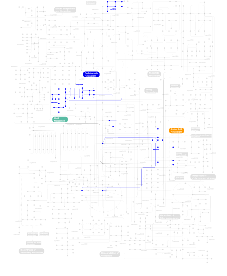
Metabolism (metabolic pathways involving proteins which contain this domain)
Click the image to view the interactive version of the map in iPath
This information is based on mapping of SMART genomic protein database to KEGG orthologous groups. Percentage points are related to the number of proteins with Zn_dep_PLPC domain which could be assigned to a KEGG orthologous group, and not all proteins containing Zn_dep_PLPC domain. Please note that proteins can be included in multiple pathways, ie. the numbers above will not always add up to 100%.