
cNMPCyclic nucleotide-monophosphate binding domain |
|---|
| SMART accession number: | SM00100 |
|---|---|
| Description: | Catabolite gene activator protein (CAP) is a prokaryotic homologue of eukaryotic cNMP-binding domains, present in ion channels, and cNMP-dependent kinases. |
| Interpro abstract (IPR000595): | Proteins that bind cyclic nucleotides (cAMP or cGMP) share a structural domain of about 120 residues [ (PUBMED:14638413) (PUBMED:10550204) (PUBMED:1710853) ]. The best studied of these proteins is the prokaryotic catabolite gene activator (also known as the cAMP receptor protein) (gene crp) where such a domain is known to be composed of three alpha-helices and a distinctive eight-stranded, antiparallel beta-barrel structure. There are six invariant amino acids in this domain, three of which are glycine residues that are thought to be essential for maintenance of the structural integrity of the beta-barrel. cAMP- and cGMP-dependent protein kinases (cAPK and cGPK) contain two tandem copies of the cyclic nucleotide-binding domain. The cAPK's are composed of two different subunits, a catalytic chain and a regulatory chain, which contains both copies of the domain. The cGPK's are single chain enzymes that include the two copies of the domain in their N-terminal section. Vertebrate cyclic nucleotide-gated ion-channels also contain this domain. Two such cations channels have been fully characterised, one is found in rod cells where it plays a role in visual signal transduction. |
| Family alignment: |
There are 149838 cNMP domains in 136080 proteins in SMART's nrdb database.
Click on the following links for more information.
- Evolution (species in which this domain is found)
-
Taxonomic distribution of proteins containing cNMP domain.
This tree includes only several representative species. The complete taxonomic breakdown of all proteins with cNMP domain is also avaliable.
Click on the protein counts, or double click on taxonomic names to display all proteins containing cNMP domain in the selected taxonomic class.
- Cellular role (predicted cellular role)
-
Binding / catalysis: cAMP-binding, cGMP-binding
- Literature (relevant references for this domain)
-
Primary literature is listed below; Automatically-derived, secondary literature is also avaliable.
- Shabb JB, Poteet CE, Kapphahn MA, Muhonen WM, Baker NE, Corbin JD
- Characterization of the isolated cAMP-binding B domain of cAMP-dependent protein kinase.
- Protein Sci. 1995; 4: 2100-6
- Display abstract
A 14.4-kDa cAMP-binding fragment was generated during bacterial expression and purification of recombinant bovine cAMP-dependent protein kinase type I alpha regulatory subunit (RI alpha). The full-length RI alpha from which the fragment was derived contained a point mutation allowing its B domain to bind both cAMP and cGMP with high affinity while leaving its A domain highly cAMP selective. The NH2 terminus of the fragment was Ser-252, indicating that it encompassed the entire predicted B domain. Although the [3H]cAMP and [3H]cGMP exchange rates of the isolated B domain were increased relative to the B domain in intact RI alpha, the [3H]cAMP exchange rate was comparable to that of the B domain of full-length RI alpha containing an unoccupied A domain. A plasmid encoding only the isolated B domain was overexpressed in Escherichia coli, and a monomeric form of the B domain was purified that had identical properties to the proteolytically generated fragment, indicating that all of the elements for the high-affinity cAMP-binding B domain are contained within the 128 amino acid carboxyl terminus of the R subunit. Prolonged induction of the B domain in E. coli or storage of the purified protein resulted in the formation of a dimer that could be reverted to the monomer by incubation in 2-mercaptoethanol. Dimerization caused an approximate fivefold increase in the rate of cyclic nucleotide exchange relative to the monomer. The results show that an isolated cAMP-binding domain can function independently of any other domain structures of the R subunit.
- Schultz SC, Shields GC, Steitz TA
- Crystal structure of a CAP-DNA complex: the DNA is bent by 90 degrees.
- Science. 1991; 253: 1001-7
- Display abstract
The 3 angstrom resolution crystal structure of the Escherichia coli catabolite gene activator protein (CAP) complexed with a 30-base pair DNA sequence shows that the DNA is bent by 90 degrees. This bend results almost entirely from two 40 degrees kinks that occur between TG/CA base pairs at positions 5 and 6 on each side of the dyad axis of the complex. DNA sequence discrimination by CAP derives both from sequence-dependent distortion of the DNA helix and from direct hydrogen-bonding interactions between three protein side chains and the exposed edges of three base pairs in the major groove of the DNA. The structure of this transcription factor--DNA complex provides insights into possible mechanisms of transcription activation.
- Weber IT, Steitz TA, Bubis J, Taylor SS
- Predicted structures of cAMP binding domains of type I and II regulatory subunits of cAMP-dependent protein kinase.
- Biochemistry. 1987; 26: 343-51
- Display abstract
The mammalian cAMP-dependent protein kinases have regulatory (R) subunits that show substantial homology in amino acid sequence with the catabolite gene activator protein (CAP), a cAMP-dependent gene regulatory protein from Escherichia coli. Each R subunit has two in-tandem cAMP binding domains, and the structure of each of these domains has been modeled by analogy with the crystal structure of CAP. Both the type I and II regulatory subunits have been considered, so that four cAMP binding domains have been modeled. The binding of cAMP in general is analogous in all the structures and has been correlated with previous results based on photolabeling and binding of cAMP analogues. The model predicts that the first cAMP binding domain correlates with the previously defined fast dissociation site, which preferentially binds N6-substituted analogues of cAMP. The second domain corresponds to the slow dissociation site, which has a preference for C8-substituted analogues. The model also is consistent with cAMP binding in the syn conformation in both sites. Finally, this model has targeted specific regions that are likely to be involved in interdomain contacts. This includes contacts between the two cAMP binding domains as well as contacts with the amino-terminal region of the R subunit and with the catalytic subunit.
- McKay DB, Steitz TA
- Structure of catabolite gene activator protein at 2.9 A resolution suggests binding to left-handed B-DNA.
- Nature. 1981; 290: 744-9
- Display abstract
The 2.9 A resolution crystal structure of Escherichia coli catabolite gene activator protein (CAP) complexed with cyclic AMP reveals two distinct structural domains separated by a cleft. The smaller carboxy-terminal domain is presumed to bind DNA while the amino-terminal domain is seen to bind cyclic AMP. Model building studies suggest that CAP binds to left-handed B-type DNA, contracting its major groove via two alpha-helices. It is possible that the CAP conversion of right- to left-handed DNA in a closed supercoil, is what activates transcription by RNA polymerase.
- McKay DB, Fried MG
- Crystallization and preliminary X-ray diffraction data for the cyclic AMP receptor protein of Escherichia coli.
- J Mol Biol. 1980; 139: 95-6
- Disease (disease genes where sequence variants are found in this domain)
-
SwissProt sequences and OMIM curated human diseases associated with missense mutations within the cNMP domain.
Protein Disease Potassium voltage-gated channel subfamily H member 2 (Q12809) (SMART) OMIM:152427: Long QT syndrome-2 Cyclic nucleotide-gated cation channel alpha-3 (Q16281) (SMART) OMIM:216900: Achromatopsia
OMIM:600053: Achromatopsia-2
OMIM:216900: - Metabolism (metabolic pathways involving proteins which contain this domain)
-
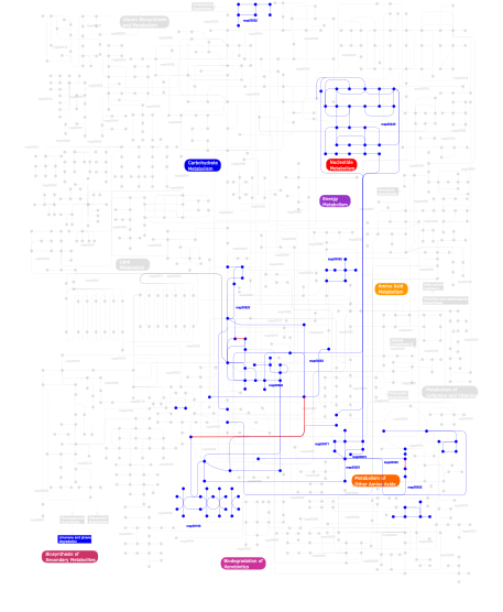
Click the image to view the interactive version of the map in iPath% proteins involved KEGG pathway ID Description 16.00 map04740 Olfactory transduction 12.44 map04210 Apoptosis 12.44 map04910 Insulin signaling pathway 8.89 map02010 ABC transporters - General 8.00  map00240
Pyrimidine metabolism 7.11 map04540 Gap junction 7.11 map04730 Long-term depression 6.67 map04670 Leukocyte transendothelial migration 3.56 map00910Nitrogen metabolism 3.56 map04720 Long-term potentiation 3.11 map00251Glutamate metabolism 3.11 map00471D-Glutamine and D-glutamate metabolism 2.22 map04010 MAPK signaling pathway 1.33 map00620Pyruvate metabolism 0.89 map00632Benzoate degradation via CoA ligation 0.44 map00562Inositol phosphate metabolism 0.44 map00190Oxidative phosphorylation 0.44 map00252Alanine and aspartate metabolism 0.44 map00903 Limonene and pinene degradation 0.44 map00330Arginine and proline metabolism 0.44 map03090 Type II secretion system 0.44 map00430Taurine and hypotaurine metabolism 0.44 map00640Propanoate metabolism This information is based on mapping of SMART genomic protein database to KEGG orthologous groups. Percentage points are related to the number of proteins with cNMP domain which could be assigned to a KEGG orthologous group, and not all proteins containing cNMP domain. Please note that proteins can be included in multiple pathways, ie. the numbers above will not always add up to 100%.
- Structure (3D structures containing this domain)
3D Structures of cNMP domains in PDB
PDB code Main view Title 1cgp 
CATABOLITE GENE ACTIVATOR PROTEIN (CAP)/DNA COMPLEX + ADENOSINE-3',5'-CYCLIC-MONOPHOSPHATE 1cx4 
CRYSTAL STRUCTURE OF A DELETION MUTANT OF THE TYPE II BETA REGULATORY SUBUNIT OF CAMP-DEPENDENT PROTEIN KINASE 1ft9 
STRUCTURE OF THE REDUCED (FEII) CO-SENSING PROTEIN FROM R. RUBRUM 1g6n 
2.1 ANGSTROM STRUCTURE OF CAP-CAMP 1hw5 
THE CAP/CRP VARIANT T127L/S128A 1i5z 
STRUCTURE OF CRP-CAMP AT 1.9 A 1i6x 
STRUCTURE OF A STAR MUTANT CRP-CAMP AT 2.2 A 1j59 
CATABOLITE GENE ACTIVATOR PROTEIN (CAP)/DNA COMPLEX + ADENOSINE-3',5'-CYCLIC-MONOPHOSPHATE 1lb2 
Structure of the E. coli alpha C-terminal domain of RNA polymerase in complex with CAP and DNA 1ne4 
Crystal Structure of Rp-cAMP Binding R1a Subunit of cAMP-dependent Protein Kinase 1ne6 
Crystal structure of Sp-cAMP binding R1a subunit of cAMP-dependent protein kinase 1o3q 
PROTEIN-DNA RECOGNITION AND DNA DEFORMATION REVEALED IN CRYSTAL STRUCTURES OF CAP-DNA COMPLEXES 1o3r 
PROTEIN-DNA RECOGNITION AND DNA DEFORMATION REVEALED IN CRYSTAL STRUCTURES OF CAP-DNA COMPLEXES 1o3s 
PROTEIN-DNA RECOGNITION AND DNA DEFORMATION REVEALED IN CRYSTAL STRUCTURES OF CAP-DNA COMPLEXES 1o3t 
PROTEIN-DNA RECOGNITION AND DNA DEFORMATION REVEALED IN CRYSTAL STRUCTURES OF CAP-DNA COMPLEXES 1o5l 
Crystal structure of Transcriptional regulator (TM1171) from Thermotoga maritima at 2.30 A resolution 1o7f 
CRYSTAL STRUCTURE OF THE REGULATORY DOMAIN OF EPAC2 1q3e 
HCN2J 443-645 in the presence of cGMP 1q43 
HCN2I 443-640 in the presence of cAMP, selenomethionine derivative 1q5o 
HCN2J 443-645 in the presence of cAMP, selenomethionine derivative 1rgs 
REGULATORY SUBUNIT OF CAMP DEPENDENT PROTEIN KINASE 1rl3 
Crystal structure of cAMP-free R1a subunit of PKA 1run 
CATABOLITE GENE ACTIVATOR PROTEIN (CAP)/DNA COMPLEX + ADENOSINE-3',5'-CYCLIC-MONOPHOSPHATE 1ruo 
CATABOLITE GENE ACTIVATOR PROTEIN (CAP) MUTANT/DNA COMPLEX + ADENOSINE-3',5'-CYCLIC-MONOPHOSPHATE 1u12 
M. loti cyclic nucleotide binding domain mutant 1vp6 
M.loti ion channel cylic nucleotide binding domain 1wgp 
Solution structure of the cNMP-binding domain from Arabidopsis thaliana cyclic nucleotide-regulated ion channel 1zrc 
4 Crystal structures of CAP-DNA with all base-pair substitutions at position 6, CAP-ICAP38 DNA 1zrd 
4 crystal structures of CAP-DNA with all base-pair substitutions at position 6, CAP-[6A;17T]ICAP38 DNA 1zre 
4 crystal structures of CAP-DNA with all base-pair substitutions at position 6, CAP-[6G;17C]ICAP38 DNA 1zrf 
4 crystal structures of CAP-DNA with all base-pair substitutions at position 6, CAP-[6C;17G]ICAP38 DNA 1zyb 
Crystal structure of transcription regulator from Bacteroides thetaiotaomicron VPI-5482 at 2.15 A resolution 2byv 
Structure of the cAMP responsive exchange factor Epac2 in its auto- inhibited state 2cgp 
CATABOLITE GENE ACTIVATOR PROTEIN/DNA COMPLEX, ADENOSINE-3',5'-CYCLIC-MONOPHOSPHATE 2d93 
Solution structure of the cNMP_binding domain of human Rap guanine nucleotide exchange factor 6 2fmy 
CO-dependent transcription factor CooA from Carboxydothermus hydrogenoformans (Imidazole-bound form) 2gau 
Crystal structure of transcriptional regulator, Crp/Fnr family from Porphyromonas gingivalis (APC80792), Structural genomics, MCSG 2gzw 
Crystal structure of the E.coli CRP-cAMP complex 2h6b 
Crystal structure of oxidized CprK in complex with o-chlorophenolacetic acid 2h6c 
Crystal structure of reduced CprK in absence of any ligand 2hkx 
Structure of CooA mutant (N127L/S128L) from Carboxydothermus hydrogenoformans 2k0g 
Solution Structure of a Bacterial Cyclic Nucleotide-Activated K+ Channel Binding Domain in Complex with cAMP 2kxl 
Solution structure of a bacterial cyclic nucleotide-activated K+ channel binding domain in the unliganded state 2mhf 
Solution structure of the cyclic-nucleotide binding homology domain of a KCNH channel 2mng 
2MNG 2mpf 
2MPF 2n7g 
2N7G 2oz6 
Crystal Structure of Virulence Factor Regulator from Pseudomonas aeruginosa in complex with cAMP 2pqq 
Structural Genomics, the crystal structure of the N-terminal domain of a transcriptional regulator from Streptomyces coelicolor A3(2) 2ptm 
Structure and rearrangements in the carboxy-terminal region of SpIH channels 2q0a 
Structure and rearrangements in the carboxy-terminal region of SpIH channels 2qcs 
A complex structure between the Catalytic and Regulatory subunit of Protein Kinase A that represents the inhibited state 2qvs 
Crystal Structure of Type IIa Holoenzyme of cAMP-dependent Protein Kinase 2wc2 
Nmr structure of catabolite activator protein in the unliganded state 2xgx 
Crystal structure of transcription factor NtcA from Synechococcus elongatus (mercury derivative) 2xhk 
Crystal structure of transcription factor NtcA from Synechococcus elongatus bound to 2-oxoglutarate 2xko 
Crystal structure of the complex of NtcA with its transcriptional co- activator PipX 2xkp 
NtcA from Synechococcus elongatus: active and inactive 2z69 
Crystal Structure of the sensor domain of the transcriptional regulator DNR from Pseudomonas aeruginosa 2zcw 
Crystal Structure of TTHA1359, a Transcriptional Regulator, CRP/FNR family from Thermus thermophilus HB8 2zd9 
Structure of a Bacterial Cyclic-Nucleotide Regulated Ion Channel 3b02 
Crystal structure of TTHB099, a transcriptional regulator CRP family from Thermus thermophilus HB8 3beh 
Structure of a Bacterial Cyclic Nucleotide Regulated Ion Channel 3bpz 
HCN2-I 443-460 E502K in the presence of cAMP 3cf6 
Structure of Epac2 in complex with cyclic-AMP and Rap 3cl1 
M. loti cyclic-nucleotide binding domain, cyclic-GMP bound 3clp 
M. loti cyclic-nucleotide binding domain mutant 2 3co2 
Mlotik1 ion channel cyclic-nucleotide binding domain mutant 3d0s 
cAMP receptor protein from m.tuberculosis, cAMP-free form 3dkw 
Crystal Structure of DNR from Pseudomonas aeruginosa. 3dn7 
Cyclic nucleotide binding regulatory protein from Cytophaga hutchinsonii. 3dv8 
Crystal structure of a putative transcriptional regulator of the crp/fnr family (eubrec_1222) from eubacterium rectale atcc 33656 at 2.55 A resolution 3e5q 
Unbound Oxidised CprK 3e5u 
OCPA complexed CprK (C200S) 3e5x 
OCPA complexed CprK 3e6b 
OCPA complexed CprK (C200S) 3e6c 
CprK OCPA DNA Complex 3e6d 
Crystal Structure of CprK C200S 3e97 
Crystal structure of transcriptional regulator of Crp/Fnr family (YP_604437.1) from DEINOCOCCUS GEOTHERMALIS DSM 11300 at 1.86 A resolution 3etq 
X-ray structure of cysteine-free fragment of mHCN2 C-terminal region from amino acids 443-630 including C508N, C584S, and C601S mutations 3ffq 
HCN2I 443-640 apo-state 3fhi 
Crystal structure of a complex between the catalytic and regulatory (RI{alpha}) subunits of PKA 3fwe 
Crystal Structure of the Apo D138L CAP mutant 3fx3 
Structure of a putative cAMP-binding regulatory protein from Silicibacter pomeroyi DSS-3 3gyd 
Crystal structure of a cyclic nucleotide-binding domain (mfla_1926) from methylobacillus flagellatus kt at 1.79 A resolution 3h3u 
Crystal structure of CRP (cAMP receptor Protein) from Mycobacterium tuberculosis 3h3z 
Crystal structure of a putative cyclic nucleotide binding protein (spoa0323) from ruegeria pomeroyi dss-3 at 2.35 A resolution 3hif 
The crystal structure of apo wild type CAP at 3.6 A resolution. 3i54 
Crystal structure of MtbCRP in complex with cAMP 3i59 
Crystal structure of MtbCRP in complex with N6-cAMP 3idb 
Crystal structure of (108-268)RIIb:C holoenzyme of cAMP-dependent protein kinase 3idc 
Crystal structure of (102-265)RIIb:C holoenzyme of cAMP-dependent protein kinase 3iia 
Crystal structure of apo (91-244) RIa subunit of cAMP-dependent protein kinase 3iwz 
The c-di-GMP Responsive Global Regulator CLP Links Cell-Cell Signaling to Virulence Gene Expression in Xanthomonas campestris 3iyd 
Three-dimensional EM structure of an intact activator-dependent transcription initiation complex 3j4q 
Pseudo-atomic model of the AKAP18-PKA complex in a bent conformation derived from electron microscopy 3j4r 
Pseudo-atomic model of the AKAP18-PKA Complex in a linear conformation derived from electron microscopy 3kcc 
Crystal structure of D138L mutant of Catabolite Gene Activator Protein 3la2 
Crystal structure of NtcA in complex with 2-oxoglutarate 3la3 
Crystal structure of NtcA in complex with 2,2-difluoropentanedioic acid 3la7 
Crystal structure of NtcA in apo-form 3mdp 
Crystal structure of a Putative Cyclic nucleotide-binding protein (Gmet_1532) from Geobacter metallireducens GS-15 at 1.90 A resolution 3mzh 
Crystal structure of cAMP receptor protein from mycobacterium tuberculosis in complex with cAMP and its DNA binding element 3n4m 
E. coli RNA polymerase alpha subunit C-terminal domain in complex with CAP and DNA 3ocp 
Crystal structure of cAMP bound cGMP-dependent protein kinase(92-227) 3od0 
Crystal structure of cGMP bound cGMP-dependent protein kinase(92-227) 3of1 
Crystal Structure of Bcy1, the Yeast Regulatory Subunit of PKA 3ogj 
Crystal structure of partial apo (92-227) of cGMP-dependent protein kinase 3otf 
Structural basis for the cAMP-dependent gating in human HCN4 channel 3plq 
Crystal structure of PKA type I regulatory subunit bound with Rp-8-Br-cAMPS 3pna 
Crystal Structure of cAMP bound (91-244)RIa Subunit of cAMP-dependent Protein Kinase 3pvb 
Crystal structure of (73-244)RIa:C holoenzyme of cAMP-dependent Protein kinase 3qop 
Domain-domain flexibility leads to allostery within the camp receptor protein (CRP) 3r6s 
Crystal structure of GlxR transcription factor from Corynebacterium glutamicum with cAMP 3rdi 
Domain-domain flexibility leads to allostery within the camp receptor protein (CRP) 3rou 
Domain-domain flexibility leads to allostery within the camp receptor protein (CRP) 3rpq 
Domain-domain flexibility leads to allostery within the camp receptor protein (CRP) 3ryp 
Domain-domain flexibility leads to allostery within the camp receptor protein (CRP) 3ryr 
Domain-domain flexibility leads to allostery within the camp receptor protein (CRP) 3shr 
Crystal Structure of cGMP-dependent Protein Kinase Reveals Novel Site of Interchain Communication 3tnp 
Structure and Allostery of the PKA RIIb Tetrameric Holoenzyme 3tnq 
Structure and Allostery of the PKA RIIb Tetrameric Holoenzyme 3u0z 
Tetramerization dynamics of the C-terminus underlies isoform-specific cAMP-gating in HCN channels 3u10 
Tetramerization dynamics of the C-terminus underlies isoform-specific cAMP-gating in HCN channels 3u11 
Tetramerization dynamics of the C-terminus underlies isoform-specific cAMP-gating in HCN channels 3ukn 
Structure of the C-linker/CNBHD of zELK channels in C 2 2 21 space group 3ukt 
Structure of the C-linker/CNBHD of zELK channels in P1 21 1 space group 3ukv 
Structure of the C-linker/CNBHD of zELK channels in P 1 21 1 space group, crystallized in the presence of cAMP 4a2u 
CRP(CAP) from Myco. Tuberculosis, with cAMP 4ava 
Crystal structure of protein lysine acetyltransferase Rv0998 from Mycobacterium tuberculosis 4avb 
Crystal structure of protein lysine acetyltransferase Rv0998 in complex with acetyl CoA and cAMP 4avc 
Crystal structure of protein lysine acetyltransferase Rv0998 in complex with acetyl CoA and cAMP 4bh9 
A structural model of CAP mutant (T127L and S128I) in the apo state 4bhp 
A structural model of CAP mutant (T127L and S128I) in cGMP-bound state 4byy 
4BYY 4chv 
The electron crystallography structure of the cAMP-bound potassium channel MloK1 4chw 
The electron crystallography structure of the cAMP-free potassium channel MloK1 4cyd 
4CYD 4d7s 
4D7S 4d7t 
4D7T 4din 
Novel Localization and Quaternary Structure of the PKA RI beta Holoenzyme 4ev0 
Crystal Structure of Thermus thermophilus Catabolite Activator Protein 4f7z 
Conformational dynamics of exchange protein directly activated by cAMP 4f8a 
Cyclic nucleotide binding-homology domain from mouse EAG1 potassium channel 4ft8 
E. coli Catabolite Activator Protein with Cobalt and Sulfate Ligands 4hbn 
Crystal structure of the human HCN4 channel C-terminus carrying the S672R mutation 4hzf 
structure of the wild type Catabolite gene Activator Protein 4i01 
Structure of the mutant Catabolite gen activator protein V140L 4i02 
structure of the mutant Catabolite gene activator protein V140A 4i09 
structure of the mutant Catabolite gene activator protein V132L 4i0a 
structure of the mutant Catabolite gene activator protein V132A 4i0b 
structure of the mutant Catabolite gene activator protein H160L 4i2o 
The Structure of FixK2 from Bradyrhizobium japonicum 4jv4 
Crystal Structure of RIalpha(91-379) bound to HE33, a N6 di-propyl substituted cAMP analog 4jva 
Crystal Structure of RIIbeta(108-402) bound to HE33, a N6 di-propyl substituted cAMP analog 4k8f 
Structure of the heme domain of CooA from Rhodospirillum rubrum 4kg1 
cGMP-responsive diguanylate cyclase 4kl1 
HCN4 CNBD in complex with cGMP 4ku7 
Structures of PKGI Reveal a cGMP-Selective Activation Mechanism 4ku8 
Structures of PKGI Reveal a cGMP-Selective Activation Mechanism 4l11 
Structure of the C-linker/CNBHD of agERG channels 4llo 
Structure of the eag domain-CNBHD complex of the mouse EAG1 channel 4mgi 
4MGI 4mgk 
4MGK 4mgy 
4MGY 4mgz 
4MGZ 4mh0 
4MH0 4muv 
4MUV 4mx3 
Crystal Structure of PKA RIalpha Homodimer 4n9h 
4N9H 4n9i 
4N9I 4nvp 
Structure of the cyclic nucleotide-binding domain of HCN4 channel complexed with 7-CH-cAMP 4off 
4OFF 4ofg 
4OFG 4oll 
cAMP-binding acyltransferase from Mycobacterium smegmatis 4onu 
cAMP-binding acyltransferase from Mycobacterium smegmatis, E234A mutant 4orf 
cAMP-binding acyltransferase from Mycobacterium smegmatis, mutant R95K 4qx5 
4QX5 4qxk 
4QXK 4r8h 
4R8H 4rz7 
4RZ7 4wbb 
4WBB 4x6q 
4X6Q 4x6r 
4X6R 4z07 
4Z07 5bv6 
5BV6 5c6c 
5C6C 5c8w 
5C8W 5ciz 
5CIZ 5cvr 
5CVR 5d1i 
5D1I 5dyk 
5DYK 5dyl 
5DYL 5dzc 
5DZC 5e16 
5E16 5e44 
5E44 5f0a 
5F0A 5fet 
5FET 5i2d 
5I2D 5j3u 
5J3U 5jon 
5JON 5k7l 
5K7L 5k8s 
5K8S 5kbf 
5KBF 5khg 
5KHG 5khh 
5KHH 5khi 
5KHI 5khj 
5KHJ 5khk 
5KHK 5t3n 
5T3N - Links (links to other resources describing this domain)
-
PFAM cNMP_binding INTERPRO IPR000595